Download
1 / 1
10 likes | 148 Views
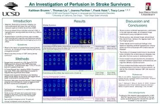
An Investigation of Perfusion in Stroke Survivors. Kathleen Brumm 1 , Thomas Liu 2 , Joanna Perthen 2 , Frank Haist 2 , Tracy Love 1, 2, 3 1 SDSU/ UCSD Joint Doctoral Program in Language and Communicative Disorders; 2 University of California, San Diego; 3 San Diego State University.

E N D
An Investigation of Perfusion in Stroke Survivors Kathleen Brumm 1, Thomas Liu 2, Joanna Perthen 2, Frank Haist 2, Tracy Love 1, 2, 3 1 SDSU/ UCSD Joint Doctoral Program in Language and Communicative Disorders; 2 University of California, San Diego; 3 San Diego State University Results Discussion and Conclusions Introduction • Magnetic Resonance Perfusion Imaging is a neuroradiological technique that can directly measure regional cerebral blood flow (rCBF) • Previous research has indicated reduced rCBF, or hypoperfusion, among stroke survivors (e.g. Hillis et al., 2001) • Hypoperfusion in stroke survivors may correlate more strongly with cognitive and behavioral deficits than standard anatomical neuroimaging (Love et al., 2002) Stroke Survivor R L R L • Stroke survivor exhibited hypoperfusion, particularly in the peri-lesional areas, as compared to age-matched and young unimpaired controls • Stroke survivor also showed longer arterial blood flow transit times, between the tagging region and the imaging regions • These results confirm previous findings in the literature regarding cerebral hypoperfusion among stroke survivors and people with cerebrovascular diseases • The finding of increased transit time for the arterial bolus indicates that cerebrovascular diseases may also alter the time course of cerebral perfusion • Future research should examine the correlations between cerebral hypoperfusion and behavioral and cognitive deficits in stroke survivors • Future studies should also explore the stability of cerebral perfusion over time in stroke survivors, as well as the correlation between CBF and neuronal plasticity msec msec L L L • Questions • What is the nature of hypoperfusion among stroke survivors, as compared to age-matched and young unimpaired controls? • What is the time course of rCBF among stroke survivors? Bolus Widths Transit delay • Transit Delays and Bolus Widths: Stroke Survivor, 45 years old, 5 years post-stroke • Representative areas of long transit delays and hypoperfusion are indicated by the yellow arrow; anatomical imaging shown at bottom right • Transit Delays (Mean and SD) • Bolus Widths (Mean and SD) Age-Matched Control R L R L Methods msec msec • Images were acquired on a GE Signa EXCITE 3Tesla scanner using an ASL FAIR sequence (TR=2500ms, 16 slices (5mm skip 1 mm), FOV=22)and an 8 gradient head coil • A Pulsed Arterial Spin Labeling (PASL) Procedure was used; PASL uses radiofrequency pulses to magnetically tag arterial blood water • Images were acquired following a post-labeling delay (transit delay) during which the tagged blood water is delivered to cerebral areas • Perfusion data were acquired across 8 transit delays (ranging from 300msec to 3 sec) to measure both the time course and localization of rCBF Bolus Widths Transit delay • Transit Delays (Mean and SD) • Bolus Widths (Mean and SD) Transit Delays and Bolus Widths: Age-matched control, 39 years old References Young Unimpaired Control Hillis, A. et al. (2001). Hypoperfusion of Wernicke’s Area predicts severity of semantic deficit in acute stroke. Annals of Neurology, 50 (5), 561-566. Love, T., Swinney, D., Wong, E., Buxton, R. (2002). Perfusion imaging and stroke: A more sensitive measure of the brain bases of cognitive deficits. Aphasiology, 16 (9), 873-883. R L R L • Participants • Participants were screened for a negative history of drug or alcohol abuse • Stroke survivor (45 years old, 5 years post-stroke): damage to left anterior cortical hemisphere; single unilateral stroke, no history of other neurological impairment • Age-matched unimpaired control, 39 years old • Younger unimpaired control, 23 years old msec msec Acknowledgements Funding for this research was supported by a NIH-NIDCD Training Grant T3 DC007361 to the first author and NIH Grants R01 DC006739 and R01 DC03681 to the last author Bolus Widths Transit delay • Transit Delays (Mean and SD) • Bolus Widths (Mean and SD) Transit Delays and Bolus Widths: Young unimpaired control, 23 years old