Download
1 / 30
300 likes | 827 Views
HEPATOBILIARY IMAGING. Presented by Yang Shiow-wen 11/26/2001. Hepatobiliary Imaging. The function of the biliary tree and gall bladder A "HIDA" scan or a "DISIDA" scan. Hepatobiliary Imaging. Performed with a variety of compounds that share the common imminodiacetate moiety.

E N D
HEPATOBILIARY IMAGING Presented by Yang Shiow-wen 11/26/2001
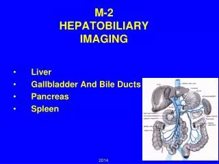
Hepatobiliary Imaging • The function of the biliary tree and gall bladder • A "HIDA" scan or a "DISIDA" scan
Hepatobiliary Imaging • Performed with a variety of compounds that share the common imminodiacetate moiety
Structures of DISIDA • Blue color: A polar component (the diacetate) • Red: A lipophilic component
Structures of DISIDA • HIDA • Little used today • DISIDA • Imaging the gall bladder better when liver function is poor
Pathways of DISIDA • Thelipophilic component : binding to hepatocyte receptors for bilirubin • Transported through the same pathways as bilirubin, except for conjugation
IDA-chelated Tc-99m • A magnification of two imminodiacetate compounds • Polar components chelated a Tc-99m molecule
Indications • Acute cholecystitis • Chronic cholecystitis • Bile leakage • Biliary atresia
Requirements for DISIDA Scan • Patient preparation: fasted for 4 hours • Radiotracer: Tc-99m IDA compounds i.v. • Imaging: serial anterior/lateral views for 60 minutes • Every 5 minutes for 30 minutes • Once at 45 minutes • Once at 1 hour • Delayed views of the gall bladder 2 hours, 4 hours, 6 hours or 24 hours after injection
Requirements for DISIDA Scan • Morphine • Injection at one hour to help force the gall bladder to fill • Water • CCK • Injection prior to the test to empty the gall bladder • Suspected chronic cholecystitis • Injection to measure how well the gall bladder empties.
Acute Cholecystitis • The most common indication • S\S • Nausea, vomiting, fever • Right upper quadrant pain post-prandially • Mild to moderate leukocytosis • Abnormal liver function test • Pain radiates to the back (scapula) • Usually blockage of the cystic duct by a gallstone
Acute Cholecystitis • If hepatic scintigraphy reveals adequate filling of the gallbladder, acute cholecystitis is effectively excluded. • Within 30 minutes, the gallbladder fails to visualize • Wait for one whole hour • Differential diagnosis for non-visualization of the gallbladder • Relaxation of the sphincter of Oddi • Inject morphine (3-5 milligrams) and continue the study for another half an hour
Non-Visualization of Gallbladder Negative study– after injection of morphine
Chronic Cholecystitis • Ultrasound is the primary modality of choice • S\S • Usually having gall stones • The cystic duct is not blocked • More chronic pain • Delayed visualization of the gall bladder • Biliary dyskinesia in response to administration of CCK
Bile leaks • Most appropriate non-invasive imaging technique for evaluation of bile leaks • Sensitivity: 87%, Specificity: 100% (2-3 ml of labeled bile) • Radiopharmaceutical activity • In an extrahepatic and extraluminal location • More intense with time • Differentiating intraluminal activity from a leak • Ingestion of water • Standing views in addition to anterior oblique views
No Excretion from Liver • No excretion up to 6 hours • This pattern is commonly seen in • Ascending cholangitis • Pancreatitis • Hepatitis
Pseudo Gallbladder Radionuclide in C-loop of the Duodenum
Pseudo Gallbladder Disappear after ingestion of water
References • http://www.vh.org/Providers/Lectures/IROCH/BiliaryNucs/BiliaryNucs.html (Virtual Hospital) • Chapter 38, Hepatobiliary Imaging, Darlene Fink-Bennett, P759-770
The End Thank for Your Attention !