Download
1 / 46
470 likes | 623 Views
Minerals . Dr. nasim. Mineral (cont.). 2 types Macro – Mineral Micro – Mineral . Macro – Mineral. Requirement more than 100mg per day. Example . Sodium Potassium Calcium Magnesium Chloride. N a +. Sodium is the principal cation of extra cellular fluid.

E N D
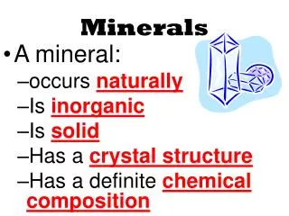
Minerals Dr. nasim
Mineral (cont.) • 2 types • Macro – Mineral • Micro – Mineral
Macro – Mineral • Requirement more than 100mg per day. Example. • Sodium • Potassium • Calcium • Magnesium • Chloride
Na+ • Sodium is the principal cation of extra cellular fluid. • It is found in all types of foods. • (RDA) is 5-10 gms. • It is excreted in the urine. • The concentrations are maintained by Aldosterone.
K+ • Potassium is intracellular cation; daily requirement is 1 gm/day. Its excretion is through kidney, • linked to sodium excretion.
Finctions of Na+ and K+ • maintains ECF balance. • Nerve conduction • Muscle contraction • Sodium is exchanged with Hydrogen in renal tubules to acidify urine. • Sodium pump keeps sodium in far higher concentration outside the cell ,create resting membrane potential. • Sodium and Potassium maintain the degree of hydration of plasma proteins, and there by viscosity of blood. • Potassium is important for functioning of cardiac muscle.
HYPER NATREMIA • HYPO NATREMIA • HYPER KALEMIA • HYPO KALEMIA
Ca+and P++++ • Mostly found in the bone. • Lesser amount found in the soft tissues, teeth and ECF.
Sources of Caand P • Milk, milk products, green leafy vegetables are rich in calcium. • Phosphate is widely distributed in nature.
Calcium: RDA 500mg for adults and 1200mg for children, 1500mg for post-menopausal women.
Absorption Increased by • cidic pH solubilizes Calcium salts, promote absorption. • High protein diet favors absorption • Vitamin D • PTH, Calcitonin • Normal blood concentration is critically maintained at 9-11 mg %
Absorption Decreased by • high fiber diet, oxalates • Glucocorticoids
Functions • Calcification of bones and teeth. • blood coagulation • Neuromuscular transmission. • Muscle contraction • Acts as secondary messenger in hormone action
P • Phosphorus: Dietary sources are cheese, milk, nuts. Eggs and organ meats. • Absorption and regulation is similar to that of Calcium
functions • Constituent of bone and teeth • Needed for the synthesis of energy rich molecules like ATP and Creatin phosphate. • It forms Phosphate buffer in blood. • Constituent of phospholipids, biomolecules and coenzymes (TPP)
Trace elements • Daily requirements of some elements is very veryless.
Iron • In body it is found in • Haemoglobin • Myoglobin • Ferritin • Hemosiderin • Transferrine • cytochromes
RDA is 10-20mgs. • Sources are meat, fish, eggs, cereals & green leafy vegetables. • Milk is deficient in Iron.
Absorption is through intestinal mucosa • It combines with intracellular binding protein Apoferritin to ferritin. Almost 300 ferric ions can bind to one molecule of apoferritin.. • For transport, free iron binds to Apo transferrin, in blood to form transferrin.It is the major • transport form of iron. It also prevents toxicity of free iron.
hemosidrosis • Excessive binding of iron causes denaturation of ferritin molecule. It undergoes aggregation, to form hemosiderin. • Mobilization of iron from hemosiderin is very slow. accumulation of hemosiderin is known as hemosiderosis.
Massive deposits of hemosiderin in tissues lead to hemachromatosis.
Harmful effect of hemachromatosis: • Damage to: • liver, • pancreas, it damages β cells, result in Bronze diabetes. • skin of the patient has bronze coloration. • Oxidative damage to cardiac muscle is a biggest concern.
Causes of iron deficiency: • Reduced dietary intake. • Hemolysis • Children who are on milk diet only are prone to iron deficiency. • Chronic bleeding, irregular menstrual cycles • Peptic ulcer, piles • Hook worm infection • Repeated malarial infections.
Deficiency leads to Iron deficiency anaemia or hypochromicmicrocyticanaemia. • It is associated with low hemoglobin and ferritin
Cu++ • Humans contain around 100 mgs of copper. Liver, brain, kidney and heart are rich in copper. • Free copper is 4%, 96 % is bound to Ceruloplasmin in body. • Sources: cereals, legumes, raisins, nutsetc
Functions • Cofactor of enzymes like: • cytochromeoxidase • dopamine decarboxylase • Tyrosinase • Cyt.Coxidase • superoxide dismutase • monoamine oxidases • Tyrosyloxidase
Copper deficiency: • Causes anaemia.(Microcytic,normochromic anemia) • Failure of melanin formation because tyrosine oxidase becomes inactive.
Menke’s disease or Kinky hair syndrome: • It is fatal sex linked recessive disorder in which there is cerebral and cerebellar degeneration, • connective tissue abnormalities and kinky hair. • Both serum [Copper] and [Ceruloplasmin] is low.
Wilson’s disease: It is an Autosomal, recessive disorder. There is a decrease in the biliary • excretion of copper. Blood and tissue copper is high in these patients. • It leads to retention of copper, followed by hepato-lenticular degeneration.
Magnesium • Sources: • Vegetables • Cereals • Beans • Potatoes • Cheese • animal tissues
M • It is absorbed from the small bowel. • It is excreted through feces, urine and sweat.
functions • It is a cofactor for peptidases, ribonucleases, glycolyticenzymes • High levels depress nerve conduction, low levels may cause Tetany. • Major part is found in bones. In teeth, it is present as dentin and enamel.
Fluorine • It is solely derived from water, tea, and fish • Daily intake should not be more than 3mg. • It is absorbed by diffusion from intestine • Mostly it is found in the bones and teeth. • It is eliminated in the urine.
Functions of F • important for tooth development • prevention of Dental Caries. • promotes bone development, • increases retention of calcium and phosphate, prevent osteoporosis
Flurosis • is due to toxicity of fluoride • It damages mitochondria • Inhibit enzymes which depend on Mg, like Succinicdehydrogenase. • Protein synthesis decreases in muscle, heart, kidney, lungs, pancreas and spleen. • Collagen synthesis is adversely affected.
Iodine • Sources: • Vegetables, fruits obtained from sea shore, sea fish are rich in iodine. People who live • on hills do not get iodine from diet. They are prone to suffer from deficiency. • It is absorbed from small intestines and transported as protein complex in plasma.
Function of I • Synthesis of thyroid hormone
Zinc • Sources are liver, milk, fish, dairy products, cereals, legumes, pulses, and spinach etc. • It is absorbed in duodenum and ileum. Absorption of Zinc from the intestine • It is transported bound to a protein (α2-macroglobulin and transferrin) • RDA is 15-20mgs for adult, 3-15mgs for infants and children
Zinc is important for the activity of a number of enzymes like • Carbonic anhydrase • DNA, RNA polymerases • Release of vitamin A from liver requires Zinc. • participates in the regeneration of rhodopsin (visual cycle). • Insulin is secreted, stored as a complex of Zinc • Helps in wound healing.
Deficiency of Zinc: • Results in dwarfism and hypogonadism • Delayed sexual development • It decreases spermatogenesis in males • irregular menstrual cycles in females. • Hepatosplenomegaly
Selenium is rich in liver, kidney, finger nails. Usually plant products are good sources than • animal based diet. • It is absorbed from duodenum, transported as selenomethionine. It forms a complex with • plasma proteins for transport. In tissues, free selenium is released. • It is excreted in urine. • RDA 50-100 μg Adult
Glutathione peroxidase is a selenium dependent enzyme. • It promotes digestion, absorption of lipids and vitamin E. • It is a part of glutathione peroxidase, prevents peroxidation of PUFA in the membranes. • It helps in the retention of vitamin E in the blood. • It is a cofactor for an enzyme involved in the synthesis of thyroid hormone.
Deficiency of selenium: • • Liver cirrhosis • • Pancreatic degeneration • • Myopathy, infertility • • Failure of growth
Toxicity: • - Selenium toxicity is called Selenosis • - Toxic dose is 900micro gram/day • - It is present in metal polishes and anti-rust compounds