Download
1 / 94
2.26k likes | 4.26k Views
AL-HEDAITHY. Medical Mycology Lecture Slides. List of Contents. AL-HEDAITHY. Primary Systemic Mycoses 63 Blastomycosis 65 Histoplasmosis 67 Coccidioidomycosis 70 Paracoccidioidomycosis 73 Candidosis and Oropharyngeal 75 mycosis (For Dental Students)

E N D
AL-HEDAITHY Medical Mycology Lecture Slides
List of Contents AL-HEDAITHY • Primary Systemic Mycoses 63 • Blastomycosis 65 • Histoplasmosis 67 • Coccidioidomycosis 70 • Paracoccidioidomycosis 73 • Candidosis and Oropharyngeal 75 • mycosis (For Dental Students) • Fungal Eye Infections 83 • Selected references 89 • Basic mycology 2 • Superficial Mycoses 9 • Pityriasis versicolor 10 • Tinea Nigra 12 • Piedra 14 • Dermatophytoses 17 • Mycetoma 25 • Rhinosporidiosis 35 Lobomycosis • Phaeohyphomycosis 37 • Chromoblastomycosis 38 • Sporotrichosis 41 • Zygomycosis 43 • Aspergillosis 48 • Pneumocystosis (PCP) 51 • Candidiasis 53 • Cryptococcosis, 60 • Trichosporonosis, Geotrichosis 1
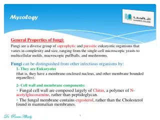
Basic Mycology AL-HEDAITHY Mycology: Mykes = Mushroom = Fungi Logos = Study of = Study of fungi Kingdom myceteae (= K. fungi) Characteristics(distinguishing features) 1) All Eukaryotic organisms 2) Heterotrophic – do not have chlorophyll (Achlorophyllous) 3) The cell is surrounded by rigid cell wall made up of chitin & complex carbohydates (Chitosan,Mannan,glucan,galactomannan) 4) Have simple structure – most of them microscopic. 5) Reproduce by spore formation sexually or asexually. Saprobic Parasitic Symbiotic 2
AL-HEDAITHY Structure of Fungi 1) Unicellular – Yeasts Examples: Saccharomyces cerevisiae True yeasts Candida albicans cells retain individuality Cells attach to each others side - by – side forming Pseudohypha 2) Filamentous – molds Examples:Aspergillus, Penicilium Rhizopus Hypha - Hyphae - Septum Yeast-like Septate hypha Nonseptate hypha (coenocytic hypha) Interwoven hyphae = Mycelium Dimorphic: Have two forms depending on change in the environmental factor like Temp., Medium and culture Vs Host Monomorphic: Only one form regardless of environment. 3
Reproduction in Fungi AL-HEDAITHY I) Asexual: Only mitotic cell division 1) Somatic Yeasts by budding Molds by hyphal fragmentation 2) Spore formation: a) Sporangiospores in sporangia b) Chlamydospores in or on hyphae c) Conidia (conidium) on hypha or on conidiophores Many types of conidia like: Blastospore, arthrospore, aleuriospore, sympodulospore…etc Asexual reproductive structures: Conidiophore Conidium Synnema Sporodochium Acervulus Pycnidium Imperfect fungi = Deuteromycetes: Do not reproduce sexually or their sexual reproduction not known e.g. Aspergillus, Fusarium, Candida 4
AL-HEDAITHY II)Sexual: Fusion, mitosis, meiosis Sexual spores: Oospore, Zygospore, Ascospore, Basidiospore Zygospore Zygomycetes e.g. Rhizopus, Mucor Ascocarp Ascus Ascospore Ascomycetes Basidiocarp Basidiospore e.g. Truffles Terfezia & Termania Basidium Gills Basidiomycetes e.g. Mushrooms & Podaxis pistillaris (Agaricus campestris) 5
AL-HEDAITHY Groups of Fungal Infections: • Superficial Mycoses • Cutaneous Mycoses • Subcutaneous Mycoses • Systemic Mycoses • Opportunistic Mycoses • Actinomycetous Infections 6
Classification of Fungi AL-HEDAITHY Taxonomy: Based on morphological, Physiological, Genetic & Molecular characteristics. Taxon = Classification rank. Kingdom: Myceteae • Binomial system Division: Amastigomycota • Latin Subdiv: Deuteromycotina Class: Deuteromycetes • Author Sub. class: Hyphomycetidae • M.canis Bodin Order: Moniliales • Primary Pathogen Family: Moniliaceae Genus: Microsporum • Opportunistic Pathogen Species: Canis Variety Strain 7
AL-HEDAITHY Classification of Fungi Kingdom Myceteae 1- Div. Gymnomycota: naked (No cell wall), phagotrophic Myxamoeba, slime molds Class Acrasiomycetes, Cl. Protosteliomycetes Cl. Myxomycetes – e.g. Dictyostelium 2- Div. Mastigomycota: flagillated, motile Cell wall, absorptive nutrition, if mold nonseptate hyphae Chytridiomycetes, Hyphochytridiomycetes Plasmodiophoromycetes, Oomycetes – e.g. Phythium, Phytophthora 3- Div. Amastigomycota: Non flagellated, Non-motile Yeasts & molds: Septate hyphae & non-septate Cl. Zygomycetes, Trichomycetes, Ascomycetes, Basidiomycetes Deuteromycetes (Fungi Imperfecti = Imperfect fungi) e.g. Aspergillus, Penicillium, Fusarium, Candida 8 Old terms: Phycomycetes, Aquatic, Lower fungi, Higher fungi
AL-HEDAITHY Superficial Mycoses • These affect the uppermost dead layers of skin or hair shaft. • They are painless and usually do not provoke the immune system • They primarily include: 1- Tinea versicolor (= Pityriasis versicolor) Brown or discolored or white patches on the skin. 2- Tinea nigra (T.n. palmaris) Dark brown or grey macular lesions – usually on palm of hand but can be on sole of foot or others. 3- Piedra Nodules of the etiologic fungus on hair shaft; a. Black piedra b. White piedra 9
Superficial Mycoses(Continued) AL-HEDAITHY Pityriasis Versicolor (= Tinea versicolor) • Brown or discolored, or white patches on skin Affect the stratum corneum The white lesions do not tan in the sun Endogenous source of infection • Etiology: Malassezia furfur It is a Yeast (= Pityrosporum orbiculare) Blastomycetidae, bipolar budding, Skin flora Lipophilic: oleic acid Or Mineral oil • Laboratory Diagnosis: Specimen is skin scrapings 10% Or 20% KOH will show short hyphal segments and round yeast cells (spaghetti & meat ball appearance) Culture on SDA and Mycobiotic medium (=SDA+ Chloramphenicol+ Cycloheximide = Actidione) with oily substance. 10
Superficial Mycoses(Continued) AL-HEDAITHY Pityriasis Versicolor Round cell Hyphal segment Scrapingsin KOH M. furfur In culture 11
AL-HEDAITHY Superficial Mycoses(Continued) Tinea nigra (= T.n. palmaris) Macular brown lesions or black stripes on palm of hand or sole of foot. Acquired by Piercing of skin with plant material in Agricultural soil. Etiology: Dematiaceous imperfect mold fungus; Phaeoannellomyces werneckii (=Exophiala werneckii) In vegetation debris In culture it produces annellospores from annellids, unicellular or two celled. Laboratory Diagnosis: Skin scrapings: - In 10% or 20% KOH will show brown septate hyphae Culture on SDA & Mycobiotic. Will grow the dematiaceous fungus. Do Lactophenol teased mount (LTM) & LPCB (Lactophenol cotton blue) for identification. 12
AL-HEDAITHY Superficial Mycoses(Continued) Tinea nigra Petri plate In KOH 3 wk old colony Conidia Phaeoannellomyceswerneckii (Exophialawerneckii) 13
Piedra AL-HEDAITHY Nodules or Encrustations on hair shaft The nodules are composed of fungal elements On scalp hair / - mustache, beard Usually in Tropics & subtropics Black piedra Assostroma with asci & ascospores Hyphal strands Firm, hard, dark brown nodules Piedraia hortae is the etiology Ascomycete – Loculoascomycetidae Cerebriform colony Hair Black piedra nodule on hair shaft Ascostroma, Asci, ascospores KOH Hair with nodule 10% -20% KOH – culture on Mycobiotic & SDA Colony of Piedraia hortaein culture 14
AL-HEDAITHY Piedra (Continued) Spores & cells White piedra Soft, brown, cream less firm Hair Etiology: Trichosporon beigelii Imperfect yeast Friable nodules Pseudohyphae, blastospores, arthrospores Culture on SDA no cycloheximide Cream – beig Yeast with wrinkled surface White piedra nodule on hair shaft KOH Arthrospore Blastospore Yeast cell Pseudohypha Culture 15
AL-HEDAITHY Treatment of Superficials • 2% salicylic acid, 3% sulfur ointments, whitfield’s ointment Ketoconazole • Cut or shave hair for Piedra & clean with mild fungicide (e.g. 1:2000 bichloride of mercury) Or apply 2% salicylic acid Or 3% sulfur ointment. Now: Nizoral shampoo for Piedra Nizoral = Ketoconazole (Benzoic acid) 16
AL-HEDAITHY Dermatophytoses • These are fungal infections of the Keratinized tissues of the body (Stratum corneum, Hair, nail) Contageous Necrotic scaley center • S/S:Skin lesion Tinea(=Ringworm) Active fungus in margin Causes itching – present anywhere in body • T.capitis in the scalp, barber’s itch The skin, hair folicles, and hair are infected. Infected hair fall-off Endothrix hair infection and Ectothrix • T.corporis : In glabrous skin • t. circinata, t.imbricata • T.pedis (=Athlete’s foot) = swimming pool itch • T.cruris: In groin = jock itch = Dhobie itch • t.unguium (nail) • t.barbae (beard) • t.manuum (hand) 17
Dermatophytoses (Continued) AL-HEDAITHY Other advanced lesions in scalp: • Kerion (pustular) • Favus (=t.favosa) with scutulum (yellow crusts) + unpleasant smell Wood’s lamp Emits filtered U.V. light, infected hair fluoresce especially microsporum spp. lesions. • Epidemiology: -Affect children + adults + males and females. More seen in school-age children -Found everywhere in the world. -Contageous – caused by primary pathogens Cats Dogs -Acquired: from infected persons and Pets -And Livestock Animals (goats, sheep, camel, cows, horses -Familial cross infection occurs 18
Dermatophytoses (Continued) AL-HEDAITHY Etiology: Dermatophytes Imperfect moniliaceous mold fungi,Primary pathogens Characteristics of dermatos: 1) Produce alkaline substance 2) Sensitive to: <20 μg/ml griseofulvin 3) Resistant to: 500 μg/ml cycloheximide 4) Have peculiar Hyphal structures like Raquet hyphae, nodular bodies, pectinate hyphae, hyphal coils (=spirals), favic chandeliers Natural Habitats: All are Keratinophilic fungi - Anthropophilic, Zoophilic, Geophilic 19
Dermatophytoses (Continued) AL-HEDAITHY • The dermatophytes are in 3 genera: Trichophyton: T.mentagrophytes in rodents,dogs,livestocks.T.violaceum in human T.verrucosum in cows. T.rubrum in human Infect skin + hair + nail Microsporum: M. canis in cats. M.audouinii in human, M. gypseum in soil Infect skin + hair, but not nail Epidermophyton floccosum in human Infect skin + nail, but not hair They reproduce asexually forming conidia by which can be identified. Perfect stage: In gymnoascaceae, Plectomycetidae ascomycetes Trichophyton in Arthroderma Microsporum in Nannizzia 20
AL-HEDAITHY -Elongated -Smooth wall -Round tip -Spindle -Rough -Pointed Macroaleuriospore (macroconidium) Microaleuriospore (Microconidium) Microsporum Trichophyton -Round tip -Club shape -Smooth wall Epidermophyton 21
Dermatophytoses (Continued) AL-HEDAITHY This is a special medium for the identification of dematophytes Dermatophyte test medium (DTM): It has pH 5.6, Antibacterial, Antifungal, and Phenol red (Amphoteric dye) In Acidic pH yellow, in alkaline red Positive DTM test is growth of the fungus and red color. It is 98% accurate. Identification Tests: • Endothrix & Ectothrix hair infection • Hair perforation test • Urease test • Pigment production in PDA & CMA media • Nutrient requirement such as – Trichophyton series Agar 1-7 22
Dermatophytoses (Continued) AL-HEDAITHY • Laboratory Diagnosis: Specimens: -Skin scrapings, Hair, Nail 20% or 10% KOH will show hyaline Septate hyphae, or spores or both. Culture on SDA and Mycobiotic medium. Identify • Treatment: • Griseofulvin Topical or systemic • Azoles Topical -Miconazole (Daktrin), Clotrimazole (Canesten), Econazole • Azoles Systemic: Itraconazole - others • Allylamines Topical, Terbinafine (Lamisil) • Tolnaftate 1% solution (=Tinactin = Tinaderm) 23
AL-HEDAITHY Dermatomycoses Other non-dermatophyte skin infections • Skin and Onychomycosis These are caused by: Candida albicans, Scytalidium, Scopulariopsis,Fusarium, Acremonium,Aspergillus,and others • Ear (Otomycosis) • Eye (oculomycosis) - Mycotic keratitis Corneal ulcer,corneal abscess - Endophthalmitis 24
AL-HEDAITHY Mycetoma (=Madura Foot) • Chronic localized subcutaneous infection that involve underlying bone later in the disease course. • The lesions are multiple abscesses. • Main symptoms/signes are cold swelling of the affected site (tumefaction), formation of sinuses that drain pus to the surface of the skin, and presence of grains. • Grains are granules (small colonies), about 1-2 mm diameter, of the etiologic agent with different color. • The commonly affected site is the foot, however, it can be in leg, thigh, hand, arm, shoulder, or head. 25
AL-HEDAITHY Mycetoma (=Madura Foot) (continued) • Infection is acquired following trauma to the skin by plant material from trees, shrubs, or vegetation debris. Thus more seen in rural areas (in farmers, Sheppards, walking bare-foot in agricultural land or city parks). • “Madura foot” referring to the first case seen in “Madura” region of India which was in the foot of that patient. • Infection is very chronic takes months to be fully established and years to deal with. It is not contagious. More seen in tropics and subtropics. • Etiologies are fungi which cause eumycotic mycetoma (Eumycetoma) or actinomycetes which cause actinomycotic mycetoma (actinomycetoma). Their natural habitats are plant materials. 26
Mycetoma (=Madura Foot) (Continued) AL-HEDAITHY • Etiology: • Eumycetoma: caused by several mold fungi. • The color of grains in this type of mycetoma is black or white. • Fungi include:Madurella, Pseudallescheria (Scedosporium), Pyrenochaeta (Pycnidia producer), Acremonium, and the ascomycetes Leptosphaeria and Neotestudina, others. • The common etiologies in Saudi Arabia and neighboring countries are: • Madurella mycetomatis causes the majority of the cases with black grains. It is imperfect dematiaceous mold with brown colonies and diffused honey – colored pigment. Produces phialoconidia from phialides, and chlamydospores Chlamydospore • Madurella grisea Another species of Madurella, similar to M.mycet. but with grey colonies. Phialoconidia Phialide Madurella mycetomatis 27
AL-HEDAITHY Mycetoma (=Madura Foot) (Continued) • Pseudallescheria boydii – causes white grain mycetoma. It is Ascomycete mold forming cleistothecia and ascospores. The imperfect of it is the moniliaceous mold: Scedosporium apiospermum which forms annelloconidia from annellids. Annelloconidia Annellid Scedosporium apiospermum 28
AL-HEDAITHY Mycetoma (=Madura Foot) (Continued) • Actinomycetoma: Caused by about 10 species of aerobic actinomycetes. • Color of grains yellow, white, yellowish-brown, pinkish – red. • Actinomycetes are filamentous higher bacteria. The filaments (very thin about 1.0 μm wide) appear as long branching, beaded, or as long rods. They are Gram-positive. • Main etiologies: • Streptomyces somaliensis- causes the majority of the cases – color of grains yellow to yellow-brown. • Actinomadura madurae – white or yellow grains. • Actinomadura pelletieri – pinkish-red grains • Nocardia brasiliensis – white grains. • N.asteroides, N. caviae, N.coeliaca – white or yellow grains. • Latter species of Nocardia usually cause Nocardiosis (which is subcutaneous, pulmonary, or brain abscess infection). 29
AL-HEDAITHY Mycetoma (=Madura Foot) (Continued) • Nocardia is acid-fast to partially acid fast when stained by Ziehl-Nelsen stain (ZN) while Streptomyces and Actinomadura are nonacid-fast. • These actinomycetes are differentiated by their decomposition pattern of casien, tyrosine, xanthine, hypoxanthine, urea, and gelatin. Also by few other biochemical tests and colony morphology(They have adherent dry colonies). See Table of characteristics. • The anaerobic actinomycete; Actinomycesisraelii causes the infection: “Actinomycosis” which is subcutaneous, cervicofacial, pulmonary, abdominal, uterine, or brain abscess infection. It also causes dental caries. The organism is filamentous and will have yellow grains (sulfer granules). 30
AL-HEDAITHY Characteristics of the main species of the above Actinomycetes Decomposition of Ca= Casien, Ty = Tyrosine, Xa = Xanthine, Hx = Hypoxanthine 31
Mycetoma (=Madura Foot) (Continued) AL-HEDAITHY • Laboratory Diagnosis: • Specimen: Visible grains, Biopsy tissue (not skin pinch), curettings of sinuses, pus, blood for serology. • First determine color of the grains – it helps identify etiology and initiate treatment. • Make histologic sections, or grinde tissue and crush grains and make smears – stain by: Hematoxylin-Eosin, Gram, ZN; if fungi do 20% KOH or Periodic acid schiff stain. Extract serum for serology. • Direct Microscopy: • Will reveal grains in tissue; • Homogenous texture actinomycetes. • Heterogenous texture fungi • Actinomycete grains will not reveal filaments easily. • Fungal grains will contain easily seen hyphae and chlamydospores. • Grains will have different morphology and color (white, black, yellow, pink, …. etc) depending on etiology. Microscope field (10X) Actinomycotic grain Eumycotic grain In tissue section 32
Mycetoma (=Madura Foot) (Continued) AL-HEDAITHY Beaded filaments • Direct microscopy of specimen from Nocardiosis and actinomycosis will show branching thin filaments by silver stain Nocardia (Gram +ve) Actinomycete filaments (Silver stain) • Culture: On SDA, Neutral – SDA, BHI-A, Blood agar and incubate at 37oC and at 25 -30oC aerobically. • For actinomycosis also culture on cooked meat medium and incubate anaerobically at 37oC • The organisms will grow slowly and may be contaminated with skin flora – purify – identify. • Serology: • Test for Antibody using known antigen from each etiologic agent. Methods used immunodiffusion (I.D), and /or counterimmunoelectrophoresis (C.I.E.) • Serology is good for Dx and monitoring treatment Actinomycete filaments fromculture (Gram +ve) 33
Mycetoma (=Madura Foot) (Continued) AL-HEDAITHY • Management: • Usually actinomycetoma respond better to treatment than eumycetoma. • Generally if bone is infected the response to treatment is poor. • Actinomycetoma: • Trimethoprim – Sulfamethoxazole (Cotrimoxazole/ Septrin) + Streptomycin sulfate • Or: Dapsone + Streptomycin sulfate • Or: Doxycycline + Cefachlor • For Actinomycosis: Penicillin G • Eumycetoma: • Ketoconazole (Nizoral) tablets or Itraconazole or voriconazole. • If drugs not effective and bone is infected Amputate the limb or debride tissue and continue Rx. • Treatment duration is long – up to years 34
Rhinosporidiosis AL-HEDAITHY • Clinical: Mucocutaneous fungal infection Sites: Nasal, Oral (Palate, epiglottis), Conjunctiva. Lesion: Polyps, Papillomas, wart-like lesions More seen in communities near swamps • Etiology: • Rhinosporidium seeberi Obligately parasitic fungus Believed to be hyphochytridiomycetes, does not grow on artificial media (e.g. SDA) But has been grown in tissue culture endospores • Laboratory Diagnosis: Specimen: Biopsy tissue Direct Microscopy: Stained sections or smears or KOH, will show spherules with endospores Culture on SDA negative spherule • Management: Cryosurgical excision of lesion-relapse common. 35
Lobomycosis AL-HEDAITHY • Clinical: Cutaneous – subcutaneous fungal infection Lesion: Keloidal – verrucoid - nodular Site: face, ear, arms, legs Chronic – localized Lacazia loboi • Etiology: (=Loboa loboi) Obligately parasitic fungus Does not grow in culture like SDA media or tissue culture • Laboratory Diagnosis:The specimen is Biopsy tissue – Direct Microscopy will show chains of cells Culture of specimen will be negative • Management: Surgical excision of lesion 36
Phaeohyphomycosis AL-HEDAITHY • Clinical: • Subcutaneous or brain Abscess caused by dematiaceous fungi • Affected site: Thigh, legs, feet, arms, ….etc, brain (cerebral) • Etiology • Dematiaceous imperfect mold fungi. Mainly: Cladosporium, Exophiala, Wangiella, Cladophialophora bantiana(Cladosporium bantianum), Ramichloridium (Rhinocladiella) mackenziei, Bipolaris, Drechslera, Rhinocladiella C.cladosporoides, E.jeanselmei, W.dermatitidis Brown Neurotropic fungi cerebral PHM as R.mack, C.bant. Pus specimen in KOH Naturally in woody plants, woods, agricultural soils • Laboratory Diagnosis: Specimens: Pus, biopsy tissue Direct Microscopy: KOH & smears brown septate hyphae Culture: On SDA & Mycobiotic – very slow growing black or grey colonies. 37
AL-HEDAITHY Chromoblastomycosis (=Chromomycosis) • Clinical: • The lesions are Hyperkeratotic, Verrucous, Pedenculus, Violaceous, Cauliflower, Initially Ulcerative, Autochthonous spread • Affected sites: extremitees, mainly feet & legs • Etiology: • Dematiaceous imperfect mold fungi in woods and woody plants. Phialophora verrucosa, Fonsecaea pedrosoi, Exophiala, Cladosporium • Laboratory Diagnosis: Specimen, Biopsy tissue • Direct Microscopy: KOH & smears brown cells with septa, Brown Muriform cells(=sclerotic bodies) • Culture: On SDA and Mycobiotic Very slow growing Dematiaceous fungi In KOH 38
AL-HEDAITHY Cladosporium Ramichloridum mackenziei Phialophora Bipolaris E.J. Blastospore Drechslera Fonsecaea pedrosoi Exophiala 39
AL-HEDAITHY Management: Phaeophypho. & Chromoblasto. • Subcutaneous: Clean surgical excision of the lesion + Antifungal • Cerebral phaeohypho: Aspiration of Pus - Antifungals • Amphotericin B, 5-Fluorocytosine (5-FC) • Azoles (e.g. Voriconazole, Posaconazole) • Caspofungin 40
Sporotrichosis AL-HEDAITHY • Clinical: • Lymphocutaneous and subcutaneous granulomatous lesions - suppurate, ulcerate. The lesions are nodules or ulcers in local lymphatics • Affected sites: extremities, joints. In agricultural communities • Etiology: Dimorphic, imperfect fungus in trees, shrubs, plant debris Sporothrix schenckii. Yeast in human tissue & at 37oC in culture. Mold in culture at room temperature with flowerettes of conidia. • Laboratory Diagnosis:Specimen:Biopsy tissue,ulcerative material Direct Microscopy: smear Finger-like yeast cells or Cigar Some are oval. Also asteroid bodies may be seen. Culture: On SDA at room temperature to grow mold, and on blood agar at 37oC to grow yeast. • Treatment: Septrin, KI 41
AL-HEDAITHY Conidium Yeast cells Asteroid body In clinical specimen and in culture at 37oC Mold in culture at room temperature Sporothrix schenckii 42
Zygomycosis AL-HEDAITHY (= Phycomycosis) Infections caused by Zygomycete fungi of the orders Mucorales & Entomophthorales. Zygomycetes are: Fast growing, moniliaceous molds, nonseptate hyphae, perfect I- Subcutaneous zygomycosis (= Entomophthoromycosis) • Clinical: Chronic localized. Subcutaneous masses, cellulitis Rhinofacial or other like Hand,Arm,Leg, thigh. Firm swelling of site with intact skin-Distortion. Acquired via nasal mucosa or insect bite, Cont. debris • Etiology: Conidiobolus coronatus, Basidiobolus ranarum, and few mucorales. These are perfect fungi that form Sporangia (conidia) and Zygospores. • Laboratory Diagnosis: Specimen: Biopsy tissue Direct microscopy: stained sections or smears will show broad non-septate hyphae with eosinophils. Culture on SDA (no antifungals), fungi will grow fast. In specimen Zygomycetes are inhibited by Cycloheximide 43 • Treatment: KI orally or KI + Ampho B or Septrin
AL-HEDAITHY Zygomycosis Sporangium Zygospore Conidiobolus coronatus Busidiobolus ranarum 44
Zygomycosis (Continued) AL-HEDAITHY II- Rhinocerebral zygomycosis (=Mucoromycosis) • Clinical: Paranasal sinusitis, orbital cellulitis Rhinofacial – orbital – craneal Usually acute, affects compromised host especially Diabetics with acidosis. Opportunistic Acquired Via nasal mucosa VERY SERIOUS – ACUTE - FATAL • Etiology: Fast growing Zygomycetes have Nonseptate hyphae of the Mucorales order maily; Rhizopus, Mucor,Absidia,Rhizomucor, others. Rhizopus arrhizus Reproduce sexually and asexually forming Sporangia with sporangiospores & Zygospores 45
Zygomycosis (Continued) AL-HEDAITHY II- Rhinocerebral zygomycosis (=Mucoromycosis) • Laboratory Diagnosis: Specimen: Biopsy tissue Direct microscopy: Stained sections or smears will show broad nonseptate hyphae Nonseptate hyphae In clinical specimen Culture on SDA (no antifungal “Cycloheximide”).The fungi will grow fast within 2-3 days. Prompt Dx & action are essential to save life • Treatment: Aggressive surgical debridement + Amphotericin B – other antifungals. III- Gastrointestinal (GI) Zygomycosis • This is a chronic zygomycete infection affecting the GI., mainly liver and intestine. The lesions are masses or abscesses in these sites. • Seen in children (6 -12 -Year old) often • Caused by Basidiobolus ranarum primarily. • Specimen: fine needle biopsy Direct Microscopy will show nonseptate hyphae & culture will grow the fungus. • Treatment: Medical with Itraconazole prognosis is good • There is mucorales G.I. Zygo which acute & Fatal. It is rare IV- Pulmonary Zygomycosis 46 • This is chronic or acute. Other aspects similar to mucoromycosis