Download
1 / 68
690 likes | 911 Views
Elbow, wrist and hand conditions. An orthopaedic overview. History. Direct Pain: Soft tissue trauma: sharp to dull aching Nerve generated: burning, lancing Joint effusion: throbbing Bone: dull boring pain Referred Pain: Joint above and below Radicular Pain: from c/s nerve root.

E N D
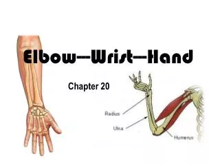
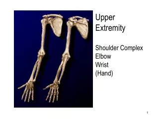
Elbow, wrist and hand conditions An orthopaedic overview
History Direct Pain: • Soft tissue trauma: sharp to dull aching • Nerve generated: burning, lancing • Joint effusion: throbbing • Bone: dull boring pain Referred Pain: Joint above and below Radicular Pain: from c/s nerve root
Age Related Incidence Young: acute trauma, overuse injury, septic arthritis, RA Older: Pathological or fragility fracture, degenerative, OA, gout “acute trauma or overuse”
Supra-condylar Fracture • Supracondylar fractures occur mostly in children and adolescents with immature skeletons. • Transcondylar fractures are more common in elderly persons with osteoporosis. • Intercondylar fractures occur in persons 40-60 years old. • Adults more likely to get radial head fracture for same MOI
Supra-condylar Fracture Most common fracture in pediatrics MOI: FOOSH extension injury (96%) Clinical: pain, swelling, bruising, deformity, NVI* Management: sling immobilizer, analgesia, ice Evacuation: X-ray, surgical consult Surgical fixation if displaced Complications: arthro-fibrosis, NVI
Condylar/ Epi-condylar Distal humerus fracture are rare MOI: axial load through elbow in varied flexion and direction produces multiple fracture patterns, high force trauma, fall, MVA Clinical: pain, swelling, deformity (altered carrying angle), guarded ROM, ASSESS for NVI Management: immobilize, analgesia, X-ray Rx: Surgical fixation to allow early mobilization Complications: arthro-fibrosis, NVI
Olecranon Fracture MOI: direct trauma to posterior aspect of elbow (fall onto olecranon) Clinical: local pain, swelling, bruising, +/- loss of active extension (avulsion of triceps) Management: immobilize, analgesia, ice, X-ray Rx: cast, ORIF (displaced) Complication: loss of extension ROM
Elbow Dislocation MOI: elbow hyper-extension via FOOSH, or valgus/supination stress during flexion Clinical: local pain, swelling, deformity, flexion contracture *Neurovascular (radial/ ulnar pulses, sensation hand) Management: immobilize, analgesia, ice, X-ray: 90 % are posterior/ postero-lateral Rx: closed reduction with procedural sedation, splinting Complications: stiffness, NVI, radial head fx, loose body
Coronoid Fracture MOI: FOOSH, avulsion or shear force from trochlea with dislocation *occur as part of a complex elbow fracture dislocation or high monteggia fracture, associated with radial head fracture, assess Neurovascular status Clinical: pain, swelling, bruising, deformity, Management: immobilize, analgesia, ice, X-ray Rx: non operative vs external fixation Complications: persistent elbow instability, heterotophic ossification
Radial Head Fracture MOI: FOOSH with elbow extended and forearm pronated (common in young adults) Clinical: Tender on palpation of radial head, decreased ROM (flex/ext), pain and blocked ROM in pron/sup Management: immobilize, analgesia, ice Evacuation for X-ray, surgical consult Rx; Undisplaced – slab and sling, early ROM Displaced – ORIF, prosthesis Complication: joint stiffness, myositis ossificans
Radial Shaft Fracture Less common than Distal radius fracture MOI: direct force (“nightstick”) or FOOSH Clinical: pain, swelling, bruising, deformity (shortening) painful ROM Management: immobilize, analgesia, ice, Evacuate: X-ray, surgical consult Rx: ORIF Complications: compartment syndrome, malunion, synostosis
What is a synostosis? • Secondary necrosis that creates an ulcer or bony synapse • Joining of one bone to another by a bony bridge
Distal Bicep’s rupture • dominant arm of middle-aged men • strong contraction of the biceps tendon against unanticipated resistance • Early surgical intervention
What three physical deficiencies would be noted with a distal biceps tear? • Inability to flex elbow with power, reduced ability to abduct the shoulder and inability to internally rotate arm. • Inability to supinate forearm with power, reduced ability to forward flex the shoulder and reduced elbow flexion power.
B! The biceps flex the elbow, help forward flex the shoulder and supinate the forearm.
Distal Radius fracture • Flexion • Extension • Fractures of the distal radius, ulna, or both, account for approximately three quarters of bony injuries of the wrist.
Extension fractures of the radius:Colles’ Fracture Transverse distal radius fx, dorsal displacement MOI: FOOSH. Lunate acts as wedge. Clinical: pain, swelling, bruising, deformity * Management: immobilize, analgesia, ice, Evacuation: X-ray, surgical consult Rx: reduction, cast or ORIF ** Complications: malunion, arthritis 60% associated with ulnar styloid fracture. 60% of ulnar styloid fractures also have associated fracture of the ulnar neck. Named after Abraham Colles (1773-1843)
Flexion fractures of the radius:Smith’s Fracture Volar tilted distal radius fracture MOI: fall onto supinated hand, roll into hyperpronation Clinical: local pain, swelling, bruising, deformity Management: immobilize, analgesia, ice Evacuate: X-ray, surgical consult Rx: reduction, cast vs ORIF (unstable) Inability to hold reduction in cast is notorious and most orthopaedic surgeons will ORIF Named after Robert William Smith
Barton’s Fracture “Push off fracture” Considered unstable Intra-articular fracture distal end of radius, involves dorsal rim w/wo dislocation MOI: extreme dorsiflexion force with pronation Clinical: pain, swelling, bruising, +/- deformity Management: immobilize, ice, analgesia Evacuate for X-ray, surgical consult Rx: ORIF if intra-articular, reduction and casting Named after John Rea Barton
Thumb fractures Bennett’s Rolando
Bennett’s/Rolando Fracture Fracture subluxation of base of 1st MC, APL attachment dislocates proximal fragment*. In Bennett’s, still articulation with carpus. In Rolando, Y-split comminution MOI: axial loading of partially flexed MC Clinical: pain, swelling, bruising, at base of thumb, CMC instability and dec ROM Management: immobilize, ice, NSAIDS, X-ray Rx: closed reduction/thumb spica, ORIF, fixation Must ORIF unstable Rolando
Boxer’s Fracture Fracture at neck of metacarpal (4th/5th) MOI: direct axial load (fist impact) Clinical: pain, swelling, bruising, depression of involved knuckle Management: immobilize, ice, NSAID, X-ray Rx: reduction, gutter splint Complications: excessive volar angulation, scissoring Some angulation is acceptable, but no twisting
Monteggia Fracture Proximal ulna fracture with dislocation of radio-capitellar joint MOI: direct trauma to post aspect of forearm, hyperpronation, fall on hyperextended elbow Clinical: pain, swelling, dec rotation of forearm, ulna angled ant, radial head dislocated ant Management: immobilization, analgesia, ice Rx: ORIF Complication: radial head fracture/ recurrent dislocation non union, NVI – posterior interosseous nerve injury
Radial Head Dislocation MOI: outstretched pronated arm, high force trauma, rarely isolated, usually complete elbow dislocation, or fracture Clinical: pain, swelling, bruising, deformity, splinting at 90 degrees Management: immobilize, analgesia, ice, X-ray* Rx: reduction** splint in supination, 90 flex Complications: compartment syndrome, recurrence, stiffness
Galeazzi Fracture Fracture of distal radial shaft with disrupted DRUJ (usually distal 1/3) MOI: fall on hand Clinical: pain, swelling, bruising, deformity Management: immobilize, analgesia, ice, X-ray Rx: ORIF of radius
Scaphoid Fracture MOI: FOOSH – produces transverse fracture at waist ( common in young men > women) Clinical: pain with wrist movement, snuff box tenderness, swelling Management: immobilize, ice, analgesia, X-ray* Rx: long arm cast with thumb spica x 4 wk, then short arm x 8 wks, vs. ext fix screw Complication: non union**, AVN ***, osteoarthritis
TFCC Injury *Triangular Fibrocartilage Complex Injuries
TFCC Injury • TFCC describes the ligamentous and cartilaginous structures that suspend the distal radius and ulnar carpus from the distal ulna. • Degeneration of the TFCC begins in the 30s and progressively increases in frequency and severity in subsequent decades. Post-fifth decade of life, no normal appearing TFCCs are seen. • Pain in ROM, DRUJ, crepitus, instability • Bracing with possible surgery
Gamekeeper’s Thumb • Pain, swelling, and/or ecchymosis of the MCP joint. Painful or weak pinch grip • A palpable mass on the ulnar aspect of the MCP joint may be obvious representing the ruptured UCL that is abnormally displaced proximally and dorsally relative to the adductor aponeurosis. • Administration of local anesthetic may be necessary to facilitate optimal examination. • A displaced avulsion fracture is a contraindication to stress testing; a nondisplaced fracture is not. • Laxity [(angulation) with thumb placed in 30° flexion] of more than 35° or laxity 15° more than the uninjured side suggests a complete rupture of the proper collateral ligament. Similar examination in extension for accessory collateral ligaments • Treatment: displaced and unstable=surgery • Stener lesion: occurs when the ruptured end of the UCL retracts and becomes abnormally displaced proximal to the adductor aponeurosis and may be palpated clinically on the ulnar side of the MCP joint, impeding proper healing.
Phalangeal Fractures MOI: direct force trauma (crush), sports injuries Clinical: pain, swelling, bruising, dec ROM, Deformity: Dorsal/Volar apex depending upon proximal or middle phalanx fracture, rotational ( scissoring effect) Management: immobilize, ice, analgesia, X-ray Rx: reduction, splinting* vs. surgical**
Phalangeal Dislocations MOI: hyper-extension force, trauma, sports injuries Clinical: MCP,IP dislocation – swelling, bruising, dec ROM, deformity (most are dorsal) Volar plates are strong stabilizing structures* Management: immobilize, analgesia, ice, X-ray** Rx: closed reduction vs surgery, splinting, physio Complications: soft tissue interposition preventing reduction, stiffness, boutonniere deformity***, unstable
Carpal Dislocations MOI: FOOSH with hand in dorsiflexion/ ulnar deviation- Lunate or perilunate dislocation Clinical: Pain, swelling, bruising, dec ROM Management: immobilize, ice, NSAIDS, X-ray Rx: r/o fracture, reduction, splinting, plastics consult Complication: chronic pain if undiagnosed, instability, AVN - lunate
Finger- Extensor Tendon Injury PIP MOI: forced flexion from active extended position (ball strike, volar dislocation) Clinical: pain at dorsal prox aspect of middle phalanx, worse with resisted ext, swelling, loss of active extension, * Management: ice, splint, NSAIDS, +/- X-ray** Rx: splinting – strict PIP extension x 6 wks Complications: boutonniere deformity
Finger- Extensor Tendon Injury DIP Most common closed finger tendon injury MOI: forced flexion of extended DIP joint (ball strike to finger tip Clinical: pain and swelling at DIP joint, flexion deformity, loss of Active extension Management: splint, ice, NSAIDS, X-ray* Rx: strict extension splinting 6-8 wks **, maintain PIP ROM Complications: mallet finger, extensor lag
Finger- Flexor Tendon Injury MOI: forced extension of actively flexed finger (Jersey Finger), most common Ring finger* Clinical: pain, swelling at volar aspect of IP joint, local tender and fullness if tendon retracts, loss of active flexion (isolate FDP and FDS ) Management: splint in current position**, ice, NSAIDS, X-ray*** Rx: early surgical repair****
Suppurative Tenosynovitis MOI: puncture wound, high pressure wound injection, disseminated GC Clinical: febrile/toxic patient, Kanavel’s cardinal signs: 1.uniform swelling 2. slight flexion position 3. pain along sheath, 4. +++pain with passive extension/ flexion* Management: early Antibiotics**, analgesia Evacuate for ongoing Rx and surgical evaluation***