Download

1 / 49
500 likes | 1.01k Views
NEUROLOGY. USMLE STEP 2 CK and 3. Neurology Outlines. Stroke (Hemorrhagic and Ischemic ) Head Trauma (see Emergency medicine and Surgery section) Neuromuscular Disease and Myopathy Demyelinating Disease Neurodegenerative Disease Dementia Spinal Cord Lesion CNS Infections

E N D
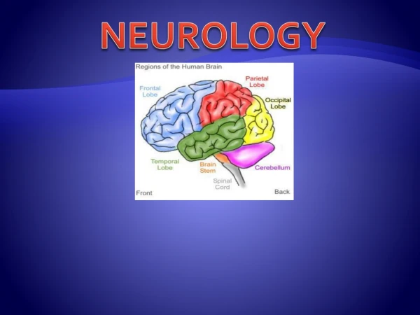
NEUROLOGY USMLE STEP 2 CK and 3
Neurology Outlines • Stroke (Hemorrhagic and Ischemic) • Head Trauma (see Emergency medicine and Surgery section) • Neuromuscular Disease and Myopathy • Demyelinating Disease • Neurodegenerative Disease • Dementia • Spinal Cord Lesion • CNS Infections • Headaches (Primary and Secondary Headache) • Seizures (Focal and Generalized) • Tremor • Brain tumor • Neurocutaneous Syndromes (SWS, NF, Tuberous Sclerosis)
PART 1: Introduction to CNS Pathphysiology • Ischemic Stroke • Emobolic • Stenotic/Thrombotic • Hemorrhagic Stroke • Subarachnoid Hemorrhage (SAH) • Intra-cerebral hemorrhage • Lacunar Stroke (MIST causing loss of direction from stroke) • Internal capsule, Thalamus, Cerebellar deficit (VBI) from small vessels of Posterior circulation • Stroke location and associated symptoms • Homonculus map • Stroke vessels and associated symptoms • Anterior Circulation (ACA, MCA) • Posterior Circulation (PICA, AICA, SCA, PCA) – brain stem and occipital lobe • Head Trauma • Epidural hematoma • Subdural hematoma • Subarachnoid Hemorrhage (SAH) • Intra-cerebral hemorrhage (not common in head trauma)
PART 2: Cerebrovascular Accident and Trauma • Ischemic Stroke • Emobolic • Stenotic/Thrombotic • Hemorrhagic Stroke • Subarachnoid Hemorrhage (SAH) • Intra-cerebral hemorrhage • Lacunar Stroke (MIST causing loss of direction from stroke) • Internal capsule, Thalamus, Cerebellar deficit (VBI) from small vessels of Posterior circulation • Stroke location and associated symptoms • Homonculus map • Stroke vessels and associated symptoms • Anterior Circulation (ACA, MCA) • Posterior Circulation (PICA, AICA, SCA, PCA) – brain stem and occipital lobe • Head Trauma • Epidural hematoma • Subdural hematoma • Subarachnoid Hemorrhage (SAH) • Intra-cerebral hemorrhage (not common in head trauma)
Case 1: Ischemic Stroke or Hemorrhagic Stroke • 58 Year old patient present with left sided weakness. He claimed that symptoms begin 20 minutes ago. Patient had a history of DM, HTN, CHF, CAD and HLP. The full stack. • What is your initial evaluation? • What is the treatment? and what would you order? • If the patient has hemorrhagic stroke, what would you expect? • If the patient has ischemic stroke, what are your treatment consideration?
PART 3: Infectious Disease of CNS • Please refer to Infectious Disease chapter • Meningitis • Encephalitis • Brain Abscess • Brain imaging choice and medical reasons behind
PART 4: Neuromuscular Disease • Myasthenia Gravis • Eaton Lambert Syndrome
PART 5: Myopathy Topics • Duchenne Muscular Dystrophy (DMD) and BMD • Polymyalgia Rheumatica (PMR) can be associated with giant cell • Inflammatory Myopathy (Dermatomyositis, Polymyositis) • Drug induced myopathy (Statin, Steroid, Amiodarone, Alcohol) • Hypothyroid Myopathy
PART 6: Demyelinating Disease CNS Demyelination • Multiple Sclerosis (MS) • Progressive Multifocal Leukoencephalopathy (PML) • AHEM/ADEM • Myelinolysis (CPM) PNS Demyelination • GullienBarre Syndrome (GBS) CNS and PNS Demyelination • Vitamin B12 • MetachromicLeukodystrophy • Krabbe’s Disease
Multiple Sclerosis • x • Etiology and Pathophysiology • x • Clinical Presentation: MC/DP • Cerebellar defects (ataxia, MLF damage) – MLF cause INO meaning diplopia • Optic neuritis (visual disturbance) • Autonomics: Bladder and bowel disturbance • Motor and Sensory disturbance • Waxing and waning or progressive • Fatigue, Spasticity (UMN) • Intention tremor and Nystagmus • Diagnosis • Initial diagnosis and Most accurate test is (MRI) • If MRI non-diagnostic, use LP to analyze CSF (oligoclonal bands) which are IgG The disorder is caused by injury or dysfunction in the medial longitudinal fasciculus (MLF), a heavily myelinated tract that allows conjugate eye movement by connecting the paramedianpontine reticular formation (PPRF) – abducensnucleus complex of the contralateral side to the oculomotor nucleus of the ipsilateral side. In young patients with bilateral INO, multiple sclerosis is often the cause.
Multiple Sclerosis • Treatment and Management • Acute Exacerbation • IV Steroid and if fail use Plasma exchange. (PO steroid worsen optic neuritis) • Relapsing and Remitting MS (use DMT: disease modifying agents) • Favor Side effect avoidance: IFN-B1a, IFN-B1b, Glatiramer [teratogens, need OCP] • Favor Efficacy: Natalizumab (Cause PML), Alentuzumab (cause ITP) • Favor Convenience: Dimethyfumarate, Teriflunomide, Fingolimod • Progressive MS • Primary Progressive: Ocrelizumab • Secondary Progressive: Continue DMT or switch DMT. Can consider add other drugs with limited evidence: MTX, Mitoxantrone, IVIG, IV steroid) • Specific Symptom Management • Fatigue – Amantadine, Modafinil, Methamphetamine, Methylphenidate, Fluorextine • Depression – SSRI/Fluroextine • Gait impairment (can’t walk properly): Dalfampridine • Spasticity – Baclofen, Tizanidine > diazepam • Trigeminal neuralgia – Carbamezapine (can mimic headache) • Bladder – Hyperactivity (Oxybutyrin), Retention bladder (Bethanechol) • ED –Sildenafil Mitoxantrone andFingolimod: Need to check for normal EF because cause cardiotoxicity • Amantadine MOA: Drug interferes with a viral protein, M2 (an ion channel), which is needed for the viral particle to become "uncoated" once it is taken inside the cell by endocytosis. • Use in Parkinson’s disease • Fatigue of MS
GBS • x • Etiology and Pathophysiology • x • Clinical Presentation: MC/DP • x • Diagnosis • x
GBS • Treatment and Management • x • Chorea Management: • x • Psychosis Management: • x • Depression: • x
PML • Progressive multifocal leukoencephalopathy (PML) is a severe demyelinating disease of the central nervous system that is caused by reactivation of the polyomavirusJC. • Asymptomatic primary infection with JC virus occurs in childhood and antibodies can be found in 85% of adults. • In most individuals, JC virus remains latent in kidneys and lymphoid organs, but, in the context of profound cellular immunosuppression, JC virus can reactivate, spread to the brain, and induce a lytic infection of oligodendrocytes, which are the CNS myelin-producing cells • Etiology and Pathophysiology • Immune-suppression (HIV or post-transplant) • IMS medications (Natalizumab for CD, MS) • HIV PML CD 4 < 200 • Clinical Presentation • Rapidly progressive and high mortality • Altered Mental status (AMS), Seizure • Motor deficit (hemiparesis/monoparesis), Limb ataxia, gait • Visual symptoms (diplopia, hemianopia) • Diagnosis • Clinical + Imaging (MRI/CT) followed by PCR JC virus DNA in CSF • Gold Standard (Brain Biopsy) • CT and MRI (T1): Hypodense (do not enhance) and lack of edema sign • MRI (T2) or FLAIR – Hyperdense on white matter (Symmetric/Asymmetric multifocal) • Treatment and Management • HIV Patient: • Optimize ART medication and aim to raise CD4+ to > 200 • Immune-suppression patient • Withdrawal IMS medications • Natalizumab using patients: • Discontinue and go for plasma exchange with mirtazepine PML image: MRI T2 Gray out, white inside No lesion in typical CVA location, no mass effect or edema seen usually
ADEM/AHEM • c • Etiology and Pathophysiology • c • Clinical Presentation • c • c • Treatment and Management • c • c
Central Pontine Myelinolysis (CPM) • c • Etiology and Pathophysiology • c • Clinical Presentation • c • c • Diagnosis: • c • c • Treatment and Management • c • c
Others Demyelinating Disease • Vitamin B12 Deficiency • Krabbe’s Disease • MetachromicLeukodystrophy
PART 7: Neurodegenerative Disease • Alzheimer’s disease (AD) and Prion’s disease, Picks Disease • Parkinson’s disease (PD) and Lewy bodies dementia (LBD) • Huntington’s disease (HD) • Cerebellar Ataxia disease with neurodegeneration • Spinocerebellar ataxia/atrophy (SCA) have many subtypes(AD inheritance) • Friedreich Ataxia • Telangiectacia ataxia • Wilson’s disease ataxia • Amyotrophic Lateral Sclerosis (ALS) • Spinal Muscular Atrophy (SMA) • Other Neurodegenerative disease: • TaySach Disease • LeshNyan Disease • SSLLAAPP=(SLAP)2, WTF HTC* Protein Misfold Autosomal Recessive 13 Neurodegeneration disease to remember
Neurodegenerative Disease • Degenerative disease are the most common form of neuro disease. • They are often depicted by progressive degeneration, death of neurons • The degeneration is typically in the gray matter • The location of the degeneration determines the type of symptoms presented • Degeneration in the cortex causes dementia • Degeneration in the basal ganglia or brain stem causes movement disorders • Parkinson’s disease • Huntington’s disease
Alzheimers Disease • Most common form of dementia depicted by slowly progressive degeneration of neurons. Constitute 50-70% of dementia. • Etiology and Pathophysiology: • Can be sporadic (90%) or hereditary (10%) • Hereditary risk can be divided into early or late onset • Early: associated with down syndrome T21, Presenilin-1/2 mutation • Late onset: associated with ApoE genotype (E2, E3, E4*): E4 worse, E2 lowest risk • Associated with decrease in Acetylcholine due to deficient of Choline Acetyltransferase which are most notable in the basal nucleus of Meynert. • Basal nucleus of Meynertinvolved in memory and cognition. • Clinical Presentation: • Short term memory loss first plus normal sensory and motor function at first • Long term memory loss and disorientation (visual spatial – lost in neighbourhood) • Loss of learnt motor or language (loss of words) • Agnoisa (difficult recognize object/spatial location), Apraxia (difficult carry out activities), Aphasia (difficulty talk) • Hallucinations, personality change, incontinence. • Diagnosis: • CT/MRI (not for diagnosis but to r/o others): Symmetric temporal and parietal atrophy. Can be diffused atrophy. • Treatment and Management: • Most common cause of death is aspiration pneumonia • Hallucination Treatment: • Memantine(NMDA blocker) • Dementia Treatment: Increase ACH with the following medication • Donepezil, Galantamine, Rivastigmine(AChE Inhibitors) – recall DRG
Parkinson’s Disease • Degeneration of dopaminergic neurons in the substantianiagra of basal ganglia. (HD is GABAnergic neurons in the Caudate nucleus of BG). • See lewy bodies and alpha-synuclein inclusions in neurons. • Etiology and Pathophysiology • Decrease dopamine, increased ACh and Increase 5HT • Clinical Presentation • Tremor (resting tremor) – pill rolling tremor • Rigidity (cogwheel rigidity) • Akinesia/bradykinesia • Postural instability (shuffling gait) • Mask-like face, Micrographia, Dementia (late stage) • Diagnosis • Clinical diagnosis, Most common cause of death is recurrent infection, fall from instability • Treatment and Management • Based on symptomatic vsNeuroprotection • Neuroprotection: L-dopa (yes), MAO-B to some extend. • Symptomatic Medications: • Dopamine precursor medications: L-dopa, Sinemet(DOC) • Dopamine agonist: Bromocriptine, Pramipexole, Rotigatone, Ropinirole, Cabergoline, Apomorphine • MAO-B inhibitors: Selegiline, Rasagiline • COMT inhibitors: Tolcapone, Entcapone • Amantadine • Anti-muscarinic: Benztrophine, Hydroxyzine
Parkinson’s Disease • Treatment for Parkinson: Medications (Symptomatic vsNeuroprotective) • Symptomatic Treatments • Dopamine Precursors (L-dopa, Sinermet = L-dopa + Carbidopa) • Dopamine Agonist (BP, RCAR) • MAO-B Inhibitors (selegiline, Rasagiline) • COMT Inhibitors (Tolcapone (tall can enter brain), Entacapone) • Amantadine (> 60 yo, so no over use of anti-cholins) • Anti-muscarinic medications (< 60 yoBenztropine, Hydroxyzine) • Neuroprotective Treatments (Disease modifying Treatments) • L-Dopa • MAO-B Inhibitors limited evidence • Dopamine agonist: • Bromocriptine • Pramipexole • Ropinirole • Rotigotine • Apomorphone • Carbegoline • MAO-B Inhibitors: So you have more Dopamine Dopamine Agonist BP high!! WHITE ROBE Black Carbegoline Roti Apomorphine
Huntington’s Disease • Autosomal Dominant disorder due to CAG repeat sequence which cause degeneration of GABAnergic neurons found in the caudate nucleus of basal ganglia • Etiology and Pathophysiology • (CAG) trinucleotide repeat expansion in the Huntingtin gene • Increase dopamine, Decrease ACh, Decrease GABA • Clinical Presentation: MC/DP • Chorea movement (rapid, involuntary, nonrepetitive or arrhythmic movement involving the face, trunk, and limbs) • Other movement disorder: Hypotonia, Hyperrefelxia, Dystonia, Bradykinesia, later can mimic parkinsonianakinetic/rigidity. • Cognitive (dementia) and Depression • Psychogenic disorder (Psychosis) • Patient present with choreiform movement disorder that can progress to dementia with depression and psychosis. • Patient can get: • Aggressive, Apathy (lack feeling and lose temper easily) • Suicidal (especially worsened with medication – tetrabenzamine) • Diagnosis • Clinical diagnosis with Genetic confirmation • MRI imaging not needed for diagnosis, more for rule out other abnormal structural findings • Will see Atrophy of caudate nucleus, dilation of lateral and 3rd ventricles.
Huntington’s Disease • Treatment and Management • Most common cause of death is suicide. • Chorea Management: • Tetrabenzamine/Deutetrabenzamine (latter less suicidal side effect) • Anti-Psychoticsmonotherapy if also have psychosis • If non-responsive to (Deu)Tetrabenzamine, use Anti-Psychotics • If severe and non-responsive to monotherapy anti-psychotics: Combo therapy (Tetrabenzamine + Anti-psychotics) • If still can’t tolerate all drug above: Keppra, Amantadine, Topiramatecan be attempted though effectiveness not established. • Psychosis Management: • Neuroleptics (Anti-psychotics: 1st or 2nd generation) • Depression: • SSRI/ >TCA • Dementia: • no real treatment • What is the MOA for tetrabenzamine? • Inhibits VMAT essential for packaging of dopamine. • Hence VMAT inhibition cause less dopamine packaged into vesicles for release.
Other Differentials • Prions • Infection by protein cause misfolding of proteins • CJD infection, see abnormal EEG, most accurate is brain biopsy, normal MRI, CSF see 14-3-3, EEG see triphasic sharp waves, seizure. • Picks disease (FTD) • Frontal temporal affected (AD is Temporal and Parietal) • Personality and behavioral change (hyperphagia/sexuality) before dementia • No movement disorder • Lewy body dementia: • Symptoms and dementia onset is very close • Less responsive to Parkinson medications.
Amyotrophic Lateral Sclerosis (ALS) • Also known as Lou Gehrig Disease. Degeneration of upper and lower motor neurons in the corticospinal/corticobulbar tract. The most common form of NGD affecting the motor system. 10% have SOD1 mutation on Chromosome 21. • Etiology and Pathophysiology: • Degeneration of UMN and LMN. TDP-43, FUS protein inclusions • Clinical Presentation: • Patient initially present Asymmetric limb weakness is the most common presentation of ALS • Can start with UMN or LMN signs. Common to start with LMN signs with atrophy and weakness of hands, fasciculation (All LMN lesion signs) • Intact: Sensory, EOM movement, Autonomics (Sexual/bladder/bowel), Intellectual unaffected, cognitive if affected affect the temporal and frontal brain. • Limb-onset: As the disease progress, man affect walking or running with tripping over/stumbling, marked by walking with a "dropped foot" which drags gently on the ground. (80%) • Bulba motor onset presentations (20%): dysarthria and dysphagia [can be due to LMN or UMN], but LMN EOM intact. • Or if arm-onset:difficulty with tasks requiring manual dexterity such as buttoning a shirt, writing, or turning a key in a lock may be experienced • Patient also can present with symptoms classic for UMN and LMN lesions • LMN of bulba(Brain stem motor neuron) innervating bulba muscles of eye are intact, but others affected (D,D) • The progressive course of ALS eventually produces one or both of the life-threatening aspects of the disease, neuromuscular respiratory failure and dysphagia. Affect muscles of respiration and swallow – Infection • Anterior motor horn degeneration (LMN lesion signs): • Flaccid Paralysis, Muscular atrophy/weakness, Fasciculation • Hypo-reflexia, negative babinski sign • Lateral corticospinal tract degeneration (UMN lesion signs) • Spastic paralysis, Increase muscle tone • Hyper-reflexia, Positive Babinski sign
Amyotrophic Lateral Sclerosis (ALS) • Complications • Many die from recurrent bouts of pulmonary infection. • Diagnosis • MRI and CSF all normal • Most accurate is EMG • Treatment and Management • Riluzole that decrease release of Glutamate • Typically 2-5 years survival after diagnosis
Other Degenerative disease • SMA • Supportive treatment • TaySach (also considered demyelinate disease) • LeshNyan Syndrome
s • Definition • x) • Etiology and Pathophysiology • x • Clinical Presentation • x • Diagnosis • x • Treatment and Management • x
x • Definition • x) • Etiology and Pathophysiology • x • Clinical Presentation • x • Diagnosis • x • Treatment and Management • x
x • Definition • x) • Etiology and Pathophysiology • x • Clinical Presentation • x • Diagnosis • x • Treatment and Management • x
x • Definition • x) • Etiology and Pathophysiology • x • Clinical Presentation • x • Diagnosis • x • Treatment and Management • x
Acute Urticaira • Definition • Urticaria (hives/wheals) is a vascular reaction of the skin. • Most cases of urticaria are self-limited and of short duration (but can recur and relapse for weeks) • Acute Urticaira (< 6 weeks); Chronic urticaria is urticarial with recurrent episodes lasting > 6 weeks) • Etiology and Pathophysiology • Increase release of histamine by basophils and mast cells – leading to leaky vessel and leaking of plasma that cause the plaque. • Sometimes plasma can go deeper into the dermis can cause angioedema • Often associated with a recent infection • Acute setting usually have 50% identifiable cause: • Food Allergy (nuts, shellfish, tomatoes, Drug Allergy (Penicillin most common, any drug possible), • Insect bite (> 24 hour lesion), Contact,Cold, Pressure, Water sunlight, exercise • Infection (acute viral syndrome, EBV, HSV, Hepatitis),. • Clinical Presentation • Transient appearance of smooth, slightly elevated papules or plaques (wheals): vascular leak hence, blench with press. • Erythematous (surrounding erythema with central halo white sometimes) • Very itchy (severe pruritus), Worse eruption and itch at night • Can occur with anaphylaxis and or angioedema (imagine the spectrum: Urticaria Angioedema anaphylaxis). • Eruption rarely lasts more than several days, it but may be recurrent over weeks. • Do not leave scar unless scratched and cause open wound. • Diagnosis • Clinical diagnosis • Treatment and Management • A short course of an oral corticosteroid (administered daily for 5-7 d, with or without a taper) or • A single dose of a long-acting injectable steroid is not usually associated with long-term sequelae and can be helpful when used for an acute episode of urticaria nonresponsive to antihistamines. • Examples of corticosteroid preparations include prednisolone, methylprednisolone, and prednisone
Patient with Seizure • 38 yo came in with history of seizure brought in by parents. What is the management for this patient? • What are the types of seizure? • What is status epilepticus? • How do you treat seizure? • What contraindication you need to be aware of.