Download
1 / 60
640 likes | 853 Views
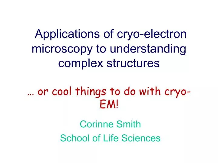
Applications of cryo-electron microscopy to understanding complex structures … or cool things to do with cryo-EM!. Corinne Smith School of Life Sciences. What cool things can we do with cryo-EM?. High resolution ‘ native-state ’ 3D protein structure

E N D
Applications of cryo-electron microscopy to understanding complex structures … or cool things to do with cryo-EM! Corinne Smith School of Life Sciences
What cool things can we do with cryo-EM? • High resolution ‘native-state’ 3D protein structure • 3D images of unique cellular structures by electron tomography (ET) • The bigger picture: Combining 3D EM with other 3D techniques • The even bigger picture: Correlating electron and light microscopy (CLEM)
Focus of the lecture: High resolution ‘native-state’ 3D protein structures • How is this achieved? – Methodology • Overview of electron tomography and CLEM
If we want to understand biological systems high resolution imaging is a powerful tool The protein folding machine, GroEL , was imaged to below 10 Å resolution using single particle cryo-EM. Because all conformations are available for analysis these structures revealed more about its mechanism than X-ray structures
X-ray crystallography NMR Cryo-electron microscopy Need crystals! – specific conditions Gives atomic resolution Single conformation seen Gives near-atomic resolution Can see dynamic processes Protein must not be too large (around 30kD) Resolution typically 7 – 30Å Crystals not always necessary Can trap intermediates Protein structure techniques
Advantages of electron microscopy • Wide range of biochemical conditions • Large, complex samples are suitable • Can work with less than 1 mg protein • Crystals and high solubility not required • All conformations available for analysis
Two main types of electron microscope • TEM transmission electron microscope • Electrons go through the sample • SEM scanning electron microscope • Electrons are reflected back off the sample
TEM vs. SEM Transmission EM Scanning EM Electron Beam Specimen Detector Detection of backscattered electrons (primary e-) and generated electrons (secondary e-) Detection of electrons behind the specimen (scattered e-) Slide courtesy of Uli Göhlke
Ground beetle • Northern Arizona University Electron Microscope Facility • http://jan.ucc.nau.edu/%7esellers/bug/99/99.html
Freeze-fracture, deep-etch rotary shadowed electron micrograph showing formation of clathrin-coated vesicles. Courtesy of John Heuser
Slide Projector Light Source Slide Condenser Lens Objective Lens Projection Screen Specimen Electron Source Transmission Electron Microscope Slide courtesy of Uli Göhlke
Stain Cryo (Holey film) Negative stain (continuous carbon) Carbon Ice
Advantages of negative stain • Heavy metal stain provides contrast • Process is quick and easy • Starting with a strongly contrasting sample allows feasibility of project to be assessed • Can obtain 3D maps of negatively stained samples
However… • With negative stain the sample is dried and could be flattened • The stain may affect the structure • Hence there is a real possibility that interesting features are artefacts
Use of ice in electron microscopy • Ice is a great fixative • Fast freezing gives vitreous ice – better for the sample and the image • A thin frozen specimen can be imaged in a specially adapted EM • The image formed is mostly a phase contrast image
Resolution of cryo-TEM 100 Å • See shape of a complex or internal structure of organelle • Subunit arrangement • Detect conformational changes • Visualise secondary structure • Atomic resolution 3 Å
Special requirements of proteins • Proteins interact with the electron beam and are destroyed by it • Proteins must be hydrated so their structure is not disturbed • Protein structure is very sensitive to the chemical environment
The Problem! Imaging proteins with an electron beam is a bit like using an atom bomb to light your living room….. …….. So you should do it indirectly!
The Solution: Low dose imaging of vitreous ice combined with image analysis
Protein structure from single protein molecules-‘single particle analysis’ • No need for crystals in order to determine protein structure! • Instead pictures of a frozen protein solution are analysed
inelastically scattered e- elastically scattered e- unmodified e- Incoming electron beam Slide courtesy of Uli Göhlke
Special characteristics of cryo-TEM image • Thin specimen scatters electrons • Interference between scattered and unscattered electrons gives phase contrast image • Image is 2D projection of original 3D object • If sufficient views are present have all the information required for 3D structure
Image formed contains a 2D projection of the original object
How is information extracted from image? • Determine the orientation of the projection with respect to the original object • Collect sufficient projections of different orientations to give complete information on object • Generate 3D model • Increase signal by averaging many projections together
Fourier Transform of Image • Fourier transform is a way of reorganising the information in an image • Performing a FT allows an image to be represented with dimensions of frequency rather than space • Result is that the nature of any repeating pattern in an image can be discovered
The FT of a sine wave Real Space FT
The Fourier transform in image processing • Any sample with a high degree of order will give a FT which can be analysed to give the positions of the ordered components (as for X-ray crystallography) • Examples are 2D crystals, helical filaments and icosahedral viruses • However, FT is also used for single particle processing
Methods to determine orientations • Angular reconstitution • Uses theory of common lines to determine relative orientations of single particles • Projection matching • Use a 3D as a template to determine orientations • Analyse FT of ordered sample • 2D protein crystals • Helical assemblies • Icosahedral virus particles (also viewed as ‘single particles’)
Resolution of reconstruction depends on: How accurately orientations are determined Access higher resolution components of image How much data you have Improve signal
Resolution v Contrast • There are two types of contrast: • Amplitude contrast gives clear images but information is lost • Phase contrast provides noisy images containing 3D information on the original object
Clathrin cages in vitro Image by John Heuser
Find Orientations 3D Reconstruction
More example of samples suitable for cryo-EM Helical assemblies Asymmetric particles 2D crystals Icosahedral viruses
2. The power of electron microscopy to see the bigger picture! • Image processing methods of frozen samples have allowed, in some cases, atomic resolution structures to be achieved • Yet we can also visualize larger objects in EM which are beyond the resolution of light microscopy
Cryo-electron tomography • Allows 3D images of complex samples to be obtained • No need for averaging procedures so can image a unique sample • Bridges the gap between light microscopy and protein structure determination methods
Cryo-electron tomography is cool – but not cool enough! • There are limitations… • The sample has to be thin, <500nm thick. This is OK for prokaryotic cells but eukaryotic cells tend to be much larger. • The electron dose should not be greater than 3000-5000 electrons nm-2 • The current geometry of image acquisition
Cryo-electron tomography of viruses • Herpes simplex virus was studied by Grünewald et al. . The virus is bound by an envelope membrane (dark blue) studded with a number of different glycoprotein spikes (yellow), enclosing the proteinaceous tegument layer (orange) and the off-centre icosahedral capsid (blue) harbouring the DNA. • (b) VV IMV was studied by Cyrklaff et al. The virion is composed of multiple layers: outer membranes (orange), enclosing the lateral bodies (red) and the dumbbell shaped core (dark blue), with a remarkably compact distribution of DNA (green) and a crystalline spike layer (light blue). • (c) Human immunodeficiency virus studied by Briggs et al. consists of an envelope (blue), and an envelope-spanning core (red) of fullerene architecture, harbouring the RNA viral genome. The density marked yellow is probably Gag-derived protein not incorporated into the core. Scale bar of 100 nm applies to all panels. Structure of complex viruses and virus-infected cells by electron cryo tomography. Curr Opin Microbiol. 2006 Aug;9(4):437-42. Grünewald K et al.
Achieving full integration of data by correlating tomograms with macromolecular structure data
Actin filaments were trapped using millisecond freezing methods and cytoplasmic protein complexes were identified using structural information
Actin and myosin structures were fitted into tomograms of rapidly frozen, contracting, insect flight muscle giving detailed information on the power stroke mechanism
Nuclear pore complexes studied in situ and in action • (a) Composite phase contrast and fluorescence micrograph of a live Dictyostelium discoideum cell, with the nucleus shown in red (mRFP-histone) and microtubules in green (GFP-tubulin). Scale bar = 5 μm. • (b) Tomographic slices from a portion of an isolated nucleus. Arrows indicate several of the nuclear pore complexes. (i) A central slice shows the nuclear envelope in cross-section. (ii) A grazing slice at the top of the nucleus shows nuclear pore complexes from the perspective of the cytoplasm. Scale bar = 200 nm.