Download
1 / 1
50 likes | 307 Views
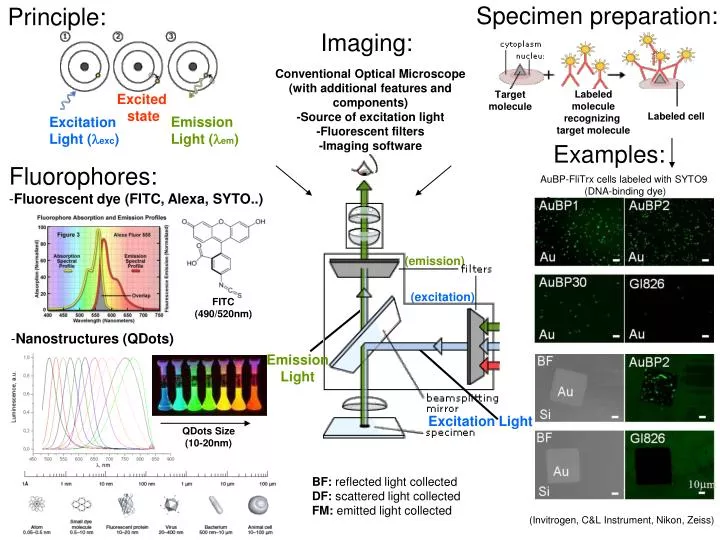
Specimen preparation:. Principle:. Imaging:. Conventional Optical Microscope (with additional features and components) -Source of excitation light -Fluorescent filters -Imaging software. Labeled molecule recognizing target molecule. Target molecule. Excited state. Labeled cell.

E N D
Specimen preparation: Principle: Imaging: Conventional Optical Microscope (with additional features and components) -Source of excitation light -Fluorescent filters -Imaging software Labeled molecule recognizing target molecule Target molecule Excited state Labeled cell Emission Light (em) Excitation Light (exc) Examples: • Fluorophores: • Fluorescent dye (FITC, Alexa, SYTO..) AuBP-FliTrx cells labeled with SYTO9 (DNA-binding dye) (emission) FITC (490/520nm) (excitation) • Nanostructures (QDots) Emission Light QDots Size (10-20nm) Excitation Light BF: reflected light collected DF: scattered light collected FM: emitted light collected (Invitrogen, C&L Instrument, Nikon, Zeiss)