Download
1 / 54
550 likes | 722 Views
Chapter 7 The Genetics of Bacteria and Their Viruses. Plasmids. Many DNA sequences in bacteria are mobile and can be transferred between individuals and among species. Plasmids are circular DNA molecules that replicate independently of the bacterial chromosome.

E N D
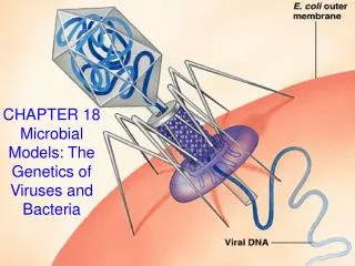
Chapter 7 The Genetics of Bacteria and Their Viruses
Plasmids • Many DNA sequences in bacteria are mobile and can be transferred between individuals and among species. • Plasmids are circular DNA molecules that replicate independently of the bacterial chromosome. • Plasmids often carry antibiotic resistance genes • Plasmids are used in genetic engineering as gene transfer vectors
F factor and Conjugation • F (fertility) factor is a conjugative plasmid transferred from cell to cell by conjugation • F factor is an episome–a genetic element that can insert into chromosome or replicate as circular plasmid • The F plasmid is a low-copy-number plasmid ~100 kb in length and is present in 1–2 copies per cell • It replicates once per cell cycle and segregates to both daughter cells in cell division
F factor and Conjugation • Conjugation is a process in which DNA is transferred from bacterial donor cell to a recipient cell by cell-to-cell contact • Cells that contain the F plasmid are donors and are designated the F+ • Cells lacking F are recipients and are designated the F– • The transfer is mediated by a tube-like structure called a pilus, formed between the cells, through which the plasmid DNA passes
Transposable Elements • Transposable elements are DNA sequences that can jump from one position to another or from one DNA molecule to another • Bacteria contain a wide variety of transposable elements • The smallest and simplest are insertion sequences, or IS elements, which are 1–3 kb in length and encode the transposase protein required for transposition and one or more additional proteins that regulate the rate of transposition
Transposable Elements • Other transposable elements in bacteria contain one or more genes unrelated to transposition that can be mobilized along with the transposable element; this type of element is called a transposon • Transposons can insert into plasmids that can be transferred to recipient cells by conjugation • Transposable elements are flanked by inverted repeats and often contain multiple antibiotic resistance genes
Figure 7.5: Cointergrate formed between two plasmids by recombination between homologous sequences present in both plasmids Figure 05: Cointegrate
Transposable Elements • Integron is a DNA element that encodes a site-specific recombinase as well as a recognition region that allows other sequences with similar recognition regions to be incorporated into the integron by recombination. • The elements that integrons acquire are known as cassettes • Integrons may acquire multiple-antibiotic-resistance cassettes, which results in the plasmid resistant to a large number of completely unrelated antibiotics • Bacteria with resistance to multiple antibiotics are an increasing problem in public health
Figure 06: Site-specific recombinase Figure 7.6: : Site-specific recombinase
Figure 07: Mechanism by which an integron sequentially captures cassettes by site-specific recombination Figure 7.7: Mechanism by which an integron sequentially captures cassettes by site-specific recombination
Bacterial Genetics • Three principal types of bacterial mutants use in bacterial genetics: • Antibiotic-resistant mutants are able to grow in the presence of an antibiotic. • Nutritional mutants are unable to synthesize an essential nutrient and thus cannot grow unless the required nutrient is supplied in the medium. Such a mutant bacterium is said to be an auxotroph. • Carbon-source mutants cannot utilize particular substances as sources of energy or carbon atoms.
Figure 09: Bacterial colonies on petri dish Figure 7.9: Bacterial colonies on petri dish Courtesy of Dr. Jim Feeley/CDC
BacterialTransformation • The process of genetic alteration by pure DNA is transformation. • Recipient cells acquire genes from DNA outside the cell. • DNA is taken up by the cell and often recombines with genes on bacterial chromosome. • Transformation may alter phenotype of recipient cells. • Bacterial transformation showed that DNA is the genetic material.
Cotransformation of Linked Genes • Cotransformation: genes located close together are often transferred as a unit to recipient cell. • Cotransformation of two genes at a frequency substantially greater than the product of the single-gene transformation frequencies implies that the two genes are close together in the bacterial chromosome. • Genes that are far apart are less likely to be transferred together • Cotransformation is used to map gene order
Conjugation • In bacterial mating, conjugation, DNA transfer is unidirectional • F factor can integrate into chromosome via genetic exchange between IS elements present in F and homologous copy located anywhere in bacterial chromosome • Cells with the F plasmid integrated into the bacterial chromosome are known as Hfr cells • Hfr: High Frequency of Recombination
Hfr • In an Hfr cell the bacterial chromosome remains circular, though enlarged ~ 2 percent by the integrated F-factor DNA • When an Hfr cell undergoes conjugation, the process of transfer of the F factor is initiated in the same manner as in an F+ cell • However, because the F factor is part of the bacterial chromosome, transfer from an Hfr cell also includes DNA from the chromosome
Hfr and Conjugation • Transfer begins within an integrated F factor and proceeds in one direction • A part of F is the first DNA transferred, chromosomal genes are transferred next, and the remaining part of F is the last • The conjugating cells usually break apart long before the entire bacterial chromosome is transferred, and the final segment of F is almost never transferred • The recipient cell remains F–
Figure 11: Integration of F Figure 7.11: Integration of F
Figure 7.12: Stages in the transfer and production of recombinants
Chromosome Mapping • It takes 100 minutes for an entire bacterial chromosome to be transferred and about 2 minutes for the transfer of F • The difference reflects the relative sizes of F and the chromosome (100 kb versus 4600 kb) • Regions in the transferred DNA may incorporate into the recipient chromosome and replace homologous regions • This results in recombinant F–cells containing one or more genes from the Hfr donor cell
Table T01: Data showing the production of recombinants when mating is interrupted at various times
Chromosome Mapping • Genes in the bacterial chromosome can be mapped by Hfr x F–mating Figure 7.13a-e: Time-of-entry mapping
Chromosome Mapping Circular genetic map of E. coli shows map distances of genes in minutes Figure 7.13f: Time-of-entry mapping
Figure 15: Formation of an F’ lac plasmid Figure 15: Formation of an F’ lac plasmid by aberrant excision of F from an Hfr chromosome
Transduction • In the process of transduction, bacterial DNA is transferred from one bacterial cell to another by a phage • A generalized transducingphage transfers DNA derived from any part of the bacterial chromosome • A specialized transducing phage transfers genes from a particular region of the bacterial chromosome.
Transduction • A generalized transducingphageP1 cuts bacterial chromosome into pieces and can package bacterial DNA into phage particles –transducing particle • Transducing particle will insert ‘transduced” bacterial genes into recipient cell by infection • Transduced genes may be inserted into recipient chromosome by homologous recombination
Transduction • A typical P1 transducing particle contains from 100 to 115 kb of bacterial DNA or about 50 genes • The probability of simultaneous transduction of both markers (cotransduction) depends on how close to each other the genes are. The closer they are, the greater the frequency of cotransduction • Cotransduction provides a valuable tool for genetic linkage studies of short regions of the bacterial genome
Figure 7.17: Demonstration of linkage of the gal and bio genes
Transduction • Specialized transducing phages transduce bacterial genes at the site of prophage insertion into the bacterial chromosome • Transduction of bacterial genes occurs by aberrant excision of viral DNA, which results in the incorporation of bacterial genes into phage chromosome
Temperate Bacteriophages • Temperate bacteriophages have two life cycles: • lytic cycle = infection that results in production of progeny phage and bacterial cell lysis • lysogeny = nonproductive viral infection results in insertion of viral DNA into bacterial chromosome • Viral DNA integration = site-specific insertion into bacterial chromosome
Lytic Cycle • The reproductive cycle of a phage is called the lytic cycle • In lytic cycle: Phage DNA enters the cell and replicates repeatedly Cell ribosomes produce phage proteins • Phage DNA and proteins assemble into new phage particles • Bacterium is split open (lysis), releasing phage progeny with parental genotypes
Figure 7.18B: Large plaques in lawn of E.coli Courtesy of CDC
Lytic Cycle • When two phage particles that have different genotypes infect a single bacterial cell, new genotypes can arise by genetic recombination • This process differs from genetic recombination in eukaryotes: • the number of participating DNA molecules varies from one cell to the next • reciprocal recombinants are not always recovered in equal frequencies from a single cell
Fine Structure of the Gene • The mutation and mapping studies of rII locus of phage T4 performed by S. Benzer provided an experimental proof to important conclusions: • Genetic exchange can take place within a gene and probably between any pair of adjacent nucleotides • The unit of mutation is an individual pair of nucleotides • Mutations are not produced at equal frequencies at all sites within a gene
Figure 7.20: Array of deletion mutations Adapted from S. Benzer, Proc. Natl. Acad. Sci. USA 47(1961): 403-426.
Figure 7.21: Genetic map Adapted from S. Benzer, Proc. Natl. Acad. Sci. USA 47(1961): 403-426
Lysogenic Cycle • All phage species can undergo a lytic cycle • Phages capable of only the lytic cycle are called virulent • The alternative to the lytic cycle is called the lysogeniccycle:no progeny particles are produced, the infected bacterium survives, and a phage DNA is transmitted to each bacterial progeny cell when the cell divides • Those phages that are also capable of the lysogenic cycle are called temperate
Lysogenic Cycle • In the lysogenic cycle, a replica of the infecting phage DNA becomes integrated into the bacterial chromosome • The inserted DNA is called a prophage, and the surviving bacterial cell is called a lysogen • Many bacterial generations, after a strain has become lysogenic, the prophage can be activated, excised from the chromosome, and the lytic cycle can begin
Bacteriophage • E. coli phage is a temperate phage capable of both lytic and lysogenic, cycles • The DNA of is a linear molecule with cohesive ends (cos) that pairing yields a circular molecule • In lysogen prophage is linearly inserted between the gal and bio genes in the bacterial DNA • The sites of integration in the bacterial and phage DNA are called the bacterial attachment site and the phage attachment site