Download
1 / 45
450 likes | 502 Views
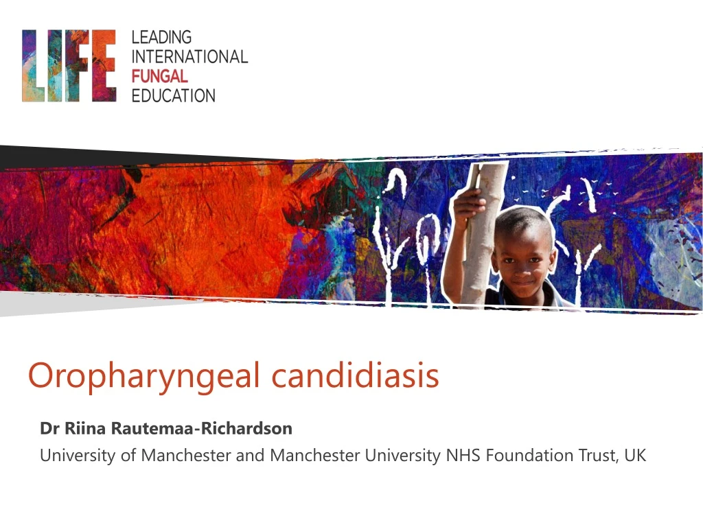
Oropharyngeal candidiasis. Dr Riina Rautemaa-Richardson University of Manchester and Manchester University NHS Foundation Trust, UK. Intended Learning Outcomes. To know the different presentations of oropharyngeal candidiasis. To know how to diagnose oropharyngeal candidiasis.

E N D
Oropharyngeal candidiasis Dr Riina Rautemaa-Richardson University of Manchester and Manchester University NHS Foundation Trust, UK
Intended Learning Outcomes To know the different presentations of oropharyngeal candidiasis To know how to diagnose oropharyngeal candidiasis To be aware of the various management approaches
What is Candida? • Over 20 species of Candida yeasts can cause infection in humans • Divided into Candida albicans and non- albicans Candida species(NAC) • Normal resident yeasts on skin, mucous membranes, GI tract without symptoms • 30-50% of healthy individuals have Candida in mouth & GI tract. • Fungal infections caused by Candida can be called ‘candidiasis’ or ‘candidosis’
Aetiology of oral candidiasis Candida albicans - most prevalent Rare causes C. glabrata (increases with increasing age) C. krusei C. tropicalis
Clinical syndromes • Candidavulvo-vaginitis • Candida perianal dermatitis • Candida balanitis • Oral candidiasis • Skin and nail infections • Sepsis and disseminated disease Immune-suppression Hypersensitivity
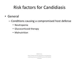
Risk factors (1) Altered normal flora: • Antibiotics, mouth rinses • Reflux (GORD) • High carbohydrate diet • Diabetes • Smoking Poor oral hygiene: • Mixed oral biofilm on non-renewing surfaces (teeth or devices such as endotracheal and nasogastric tubes)
Risk factors (2): impaired defense mechanisms SYSTEMIC: • Infancy • Diabetes • Malnutrition • Immunosuppression: • Primary • Acquired (medications, cancer, HIV) LOCAL: • Decreased saliva production • Topical corticosteroids including inhalers • Radiotherapy to head and neck
Significance of oral candidiasis • Potential for systemic invasion • Can facilitate mucosal invasion by other microbes including S. aureus • Symptom causation - nutrition, altered taste and burning sensation (impaired speech) • A risk factor for malnutrition • Development of resistance (persistent colonisation with resistant isolates) especially associated with biofilm formation
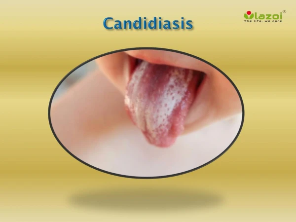
Clinical forms FOURdistinct clinical forms: • Pseudomembranous candidiasis (thrush) • Erythematous candidiasis (atrophic) • Hyperplastic candidiasis (Candida leukoplakia) • Denture stomatitis & angular cheilitis
Pseudomembranous candidiasis • Most common form • White raised lesions on tongue, mucosa, palate, tonsils • May become confluent plaques • Can be dislodged or scraped off to reveal a bleeding base • Painless
Pseudomembranous candidiasis Extensive thrush in patients who are immunocompromised note the raised soft white plaques on a red inflamed base
Erythematous candidiasis • Often associated with broad spectrum antibiotics, inhaled steroids and HIV infection • Flat red lesion usually on tongue or palate or both (“kiss lesions”) • Sore causing difficulty in feeding, especially swallowing solids • Soreness may be worsened by hot or cold liquids and spicy foods causing a burning sensation
Erythematous candidiasis Almost symmetric, asymptomatic red lesion involving the midline of the posterior dorsal tongue (known as medium rhomboid glossitis) Palatal erythematous candidiasis
Hyperplastic candidiasis • Usually on the buccal mucosa near the corner of the mouth Often has raised surface with starburst appearance; rarely on tongue (dorsal surface) • Often asymptomatic and associated with smoking or local trauma • Appearance similar to leukoplakia • Can undergo malignant transformation • Possibility of hyperplastic / dysplastic lesion with C.albicans superinfection
Angular cheilitis • Usually develops in association with other forms of oral candidiasis e.g. erythematous or denture stomatitis In older edentulous patients often associated with denture stomatitis and poor anterior oral seal leading to dribbling
Angular cheilitis • Usually develops in association with other forms of oral candidiasis e.g. erythematous or denture stomatitis Scaling, erythematous fissures at the corners of the mouth associated with infection by Candidaspp. (typically in combination with bacteria such as Staph. aureus or streptococci). In dentate patients may be associated with vitamin deficiency
Differential diagnosis • Contact dermatitis to oral hygiene products or lipsticks • All-cause cheilitis • Herpes labialis • Lichen planus
Lichen Planus Lichen planus is a chronic recurrent inflammatory mucocutaneous condition that forms plaques on the tongue and hyperkeratotic striae on the buccal mucosa that may be mistaken for candidiasis Image from DermNet New Zealand
Herpes infections Herpes labialis Herpes simplex Images from DermNet New Zealand
Aphthous ulcers Usually affect different sites to candidiasis; ulcers last 7-14 days and tend to be painful, and round or oval with wide inflammatory halo
Diagnosis • Clinical - mainly • Soreness • White plaques • Localized erythema & inflammation • Essential to rule out other causes of similar presentation • Microbiological (microscopy, culture, susceptibility testing) • Biopsy and histology for chronic hyperplastic candidiasis in order to exclude dysplastic/ malignant lesions
Microscopy: direct smear examination • Scrape the material with a spatula • Spread on glass slide then air dry • Fix with alcohol • Stain with periodic acid Schiff reagent (PAS) or potassium hydroxide (KOH) 10% • Examine under a microscope
Direct smear exam Staining with KOH PAS staining
Culture Specimen collection: - oral swab - imprint culture - oral rinse - Direct sonication of foreign body (e.g. ETT, nasogastric tube etc) Culture medium: - Sabouraud dextrose agar (SDA) Images from University of Adelaide mycology online
Biopsy • Especially for diagnosis of hyperplastic candidiasis; mainly to rule out squamous cell carcinoma and dysplasia • Histopathological exam shows epithelial parakeratosis with polymorphonuclear leukocytes in the superficial layers • PAS-stained slides show the presence of Candida hyphae
Goals of Management • Address underlying conditions / risk factors • Appropriate antifungal agents through the correct route • Consider alternative diagnosis • Prevention
How to address underlying conditions? • Mechanical / surgical disruption of biofilm • Good management of underlying disease(s) or condition(s) • Improved oral self-care • Reduce sugar intake (e.g. sweets, drinks, cakes) • Good glycemic control in diabetes • Avoid wearing dentures at night • Proper inhaled steroid use with mouth rinsing or cleaning teeth after use • Iron, vitamin B12, or folate deficiency • Smoking cessation • PPIs
Disrupting the biofilm • Drugs penetrate biofilm poorly, drug targets not active in biofilms • Azoles do not inhibit biofilm formation on surfaces and have no activity against preformed biofilms • Potential for selection of resistant strains • Mouth washes (especially those containing chlorhexidine) disrupt biofilm formation Ramageet al. Oral Surg Oral Med Oral Pathol Oral RadiolEndod 2011;111:456-60
Suggested management algorithm Rautemaa & Ramage.Crit Rev Microbiol 2011;37:328-36
Antifungal drug therapy Topical Systemic • Can be used in combination with topical antifungals • Need high concentrations and adequate saliva to reach adequate local levels (poor efficiency in dry mouth) • Preferred for immunocompromised due to high relapse rates with topical agents • Oral: fluconazole, itraconazole, posaconazole • IV: fluconazole, echinocandins, ampho B • Minimal absorption from GI tract • High local drug levels achieved • Examples: nystatin, topical miconazole • Few drug to drug interactions (miconazole interactions with warfarin and statins) • Safe in children
Options for immunocompetent infants & neonates Miconazole gel Nystatin oral suspension Oral fluconazole Neonate (up to 14 days)3–6 mg/kg, dose to be given on first day, then 3 mg/kg every 72 hours Neonate (14-28 days)3–6 mg/kg, dose to be given on first day, then 3 mg/kg every 48 hours Child (1 month–11 years)3–6 mg/kg, dose to be given on first day, then 3 mg/kg daily (max. per dose 100 mg) for 7–14 days in oropharyngeal candidiasis (max. 14 days except in severely immunocompromised patients) Neonate (BNF in UK) 1 mL 2–4 times a day treatment should be continued for at least 7 days after lesions have healed or symptoms have cleared, to be smeared around the inside of the mouth after feeds Child (1 month–1 year)1.25 ml 4 times a day treatment should be continued for at least 7 days after lesions have healed or symptoms have cleared, to be smeared around the inside of the mouth after feeds Neonate (unlicensed use) and children from BNF in UK100 000 units 4 times a day usually for 7 days, and continued for 48 hours after lesions have resolved, to be given after feeds/food Spontaneous cure in neonates by 3-8 weeks has been reported
Options for immunocompetent older children & adolescents Clotrimazole troches Dose: 1 troche (10 mg) five times daily Nystatin suspension Dose: 1ml (100,000 units/ml) every 6 hours Miconazole gel/solution Dose For Child 2–17 years (BNF, UK)2.5 ml 4 times a day treatment should be continued for at least 7 days after lesions have healed or symptoms have cleared, to be administered after meals, Retain near oral lesions before swallowing (dental prostheses and orthodontic appliances should be removed at night and brushed with gel) Fluconazole Dose: For Child 12–17 years (BNF, UK) 50 mg daily for 7–14 days in oropharyngeal candidiasis (max. 14 days except in severely immunocompromised patients)
Options for patients with immunosuppression Fluconazole is the preferred agent (in combination with topical treatment) Dose: 100-200 mg/day for 7-14 days • Child (BNF, UK)3–12 mg/kg daily (max. per dose 400 mg), commence treatment before anticipated onset of neutropenia and continue for 7 days after neutrophil count in desirable range, dose given according to extent and duration of neutropenia • Adult (BNF, UK)50–400 mg daily, commence treatment before anticipated onset of neutropenia and continue for 7 days after neutrophil count in desirable range, dose adjusted according to risk
Options for patients with immunosuppression For oral or oesophageal candidiasis that has not responded to fluconazole: • Itraconazole suspension (or capsules) Must check for drug-drug interactions and consider TDM Dose: 200-400 mg/day for 14 days (BNF, UK), by mouth using oral solution • Adult 100–200 mg twice daily for 2 weeks (continue for another 2 weeks if no response; the higher dose should not be used for longer than 2 weeks if no signs of improvement) • HIV positive or immunocompromised (BNF, UK)200 mg daily in 1–2 divided doses for 1 week (continue for another week if no response) • Other options for fluconazole-refractory disease: Oral posaconazole suspension
Response to treatment • Oral symptoms resolve in 2-5 days • Relapse is common if risk factors persist (poor oral hygiene, poorly controlled diabetes, incorrect use of inhaled steroids, untreated HIV) • Managing underlying conditions is KEY
Unresponsive oral candidiasis No response vs partial/short-lived response? Why? • Resistant organism • Alternative/additional diagnosis • Uncontrolled underlying condition (e.g. HIV / diabetes/GORD) • Failure to remove/eradicate biofilm • Chronic mucocutaneous candidiasis
Managing resistant organisms • Increasing the dose of fluconazole to 400 mg (up to 800 mg USA) for adults • Oral itraconazole or posaconazole suspension for prolonged period (up to 4 weeks) – note TDM is required • Amphotericin B oral suspension (500 mg 4x daily) may be used • IV amphotericin B or IV caspofungin
Chronic Mucocutaneous Candidiasis (CMC) • Recurrent persistent superficial candidiasis often caused by C. albicans • Genetic defect in STAT1 (AIRE) or CARD9 most common; they are IL-17 deficient • May be sporadic and not linked to family history or genetic defect • Onset in infancy for majority • Significant morbidity • Rarely disseminates
Prevention • Good oral self-care including use of chlorhexidine mouthwashes as necessary • Rinse the mouth after steroid inhaler use, use a spacer • Treat HIV • Smoking cessation • Prophylactic antifungals for secondary prevention • Avoid azoles due to resistance selection • Pulse therapy with nystatin • Probiotics and alternative agents
Summary • Primarily Candida albicans, but not always • Multiple underlying conditions of which poor oral hygiene, steroid inhalers and immunosuppression (e.g. cancer therapies and HIV) • There are 4 common clinical manifestations of oropharyngeal candidiasis, of which an inflamed mucosa is characteristic • Therapy may be with topical and/or systemic therapy depending on the patient’s immune status