Download
1 / 55
550 likes | 629 Views
The Vertiginous Patient: Diagnosing peripheral vs. central lesions. Natasha Renda 9/23/2009.

E N D
The Vertiginous Patient:Diagnosing peripheral vs. central lesions Natasha Renda 9/23/2009
“This (a patient’s complaint of dizziness) means that after an exhaustive inquiry it will still not be entirely clear what it is that the patient feels wrong or even less so why he feels it.”W.B. Matthews in Practical Neurology
Goals • Overview of anatomy and physiology of vestibulo-ocular system • Diagnosing benign positional vertigo • Diagnosing acute vestibular syndrome vs. AICA/PICA stroke • Shortcomings of the bedside examination and imaging
The vestibular system stabilizes eye position and eye movements during changes in head position to maintain focus on an intended object.
Vestibular Apparatus Ampulla Three semicircular canals: sense angular head movements.
Vestibular Apparatus Two otiliths: sense linear head movements and head (body) tilting 1. saccule aligned vertically 2. utricle aligned horizontally Macula
Vestibular Nerve • Superior division innervation: utricle, anterior and horizontal SCC’s • Inferior division innervation: saccule and posterior SCC’s
Scarpa’s Ganglion -Contains cell bodies of superior and inferior divisions -Location: internal auditory canals -Spontaneously fires at 100spikes/second :Ipsilateral angular head movements increases firing rate :Contralateral angular head movements decrease firing rate
Vascular Supply all from posterior circulation 1. Vestibular apparatus and nerve: internal auditory artery of AICA 2. Vestibular nuclei: branches of basilar and vertebrals 3. Inferior cerebellum and flocculonodular lobe: PICA/AICA
Vestibular-Ocular Reflex Moves eyes in opposite direction of head movement (rotation or linear) to maintain an image on the retina. 3 types of VOR
1. Boat pitchs up and down and veers left to right (yaw)2. Boat moves down river toward other boat3. Boat tilts left to right Want to keep looking at upcoming pontoon boat
Angular VOR: SCC Eyes move opposite to rotation of head Pitch: anterior and posterior canals Yaw: horizontal canals
2. Linear VOR: otoliths Eyes move opposite to direction of linear head movement. Riding elevator: saccule Riding train: utricle
3. Ocular tilt reflex: otilith Eyes and head move opposite to the tilt of the body. Body tilt right: elevates right eye and depresses left eye, leftward head tilt
Loss of canal function causes: Vestibular Nystagmus Example: LEFT vestibular neuritis/ left superior CN8 loss/LEFT HC and AC loss • Decreased activity from left side of causes slow phase eye movements to the left 2/2 relative excitation of firing from the right (healthy) side • Quick phases generated by burst cells (nystagmus is named according to quick phase but the slow phase points toward the deficit)
Vertigo • symptom: rotation, linear movement, tilt • Mechanism: sudden imbalance of neural activity to vestibular nucleus • Localization: labryinth, CN8, vestibular nuclei, vestibular cerebellum or central otolith pathways (OR mechanical insult to inner ear vs central pathways) • Signs: N/V, spontaneous nystagmus, postural instability
Differential using Duration of vertigo • Sub-acute attack <3 days a. vestibular neuritis b. labyrinthitis c. Meniere’s disease d. brainstem CVA: ischemic or hemorrhagic (fatal gastroenteritis) e. demyelinating 2. Chronic attack >3 days a. uncompensated unilateral vestibular defect b. bilateral hypofunction ie. ototoxictiy c. disequilibrium d. psychological 3. Episodes a. seconds: BPPV, orthostatic hypotention b. minutes: TIA, migraine, seizure, perilymphatic fistula c. hours: migraine or meniere’s disease
Alexander’s Law: peripheral vestibular nystagmus increases in intensity when the gaze is in the direction of the fast phase, and decreases in intensity when the gaze is away from the fast phase
Peripheral Nystagmus • Intensity of vestibular nystagmus and the velocity of slow phase are increased by removing fixation. • Peripheral nystagmus is unidirectional and central nystagmus is bidirectional.
To remove fixation A. Use an opthalmoscope: 1. Observe nystagmus via opthalmoscope (direction of nystagmus is inverted) 2. Cover and uncover (remove fixation) the other eye. Peripheral disorder IF direction of nystag and velocity of slow phase are increased with covering the fixating eye (removing the fixation)
To remove fixation B. Frenzel glasses
Summary of nystagmus • Pure Vertical or pure torsional: central 2. Combined vertical and torsional: peripheral or central 3. No nystagmus in vertiginous person: central
Bedside Examination • Postural instability: -Peripheral: able to walk, but uncomfortable because leaning toward side of lesion -Central: severe, unable to walk without falling 2. Romberg: -Peripheral: also fall toward side of lesion (instruct not to compensate) - Central: variable falling
Bedside Examination Central localization if other brainstem or cerebellar signs: • CN abnormalities • Motor weakness • Dysmetria • Sensory changes • Abnormal reflexes
Other Bedside Examinations Dix Hallpike test: Positive in BPPV Nystagmus begins within 30 sec and lasts <30sec. If nystagmus persists when supine, and does not occur while sitting, suspect central positional vertigo.
Other Bedside Examinations Head Impulse test: Tests the VOR: Have patient fixate on your nose. Rapidly turn patient’s head. Normal when eyes repeat fixated on nose, but will see catch up saccade in direction of your nose if abnormal. Catch up saccades in peripheral etiology but with central etiologies, test is negative.
Other Bedside Examinations Smooth Pursuit: Slowly follow moving target horizontally and vertically without moving their head - parietal-occipital cortex, pons, cerebellum cause catch-up saccades
Further Non-bedside Evaluations • Electronystagmography • Rotary chair • Caloric test • Posturography • Audiometry • Brainstem evoked potentials
Case 1: 55year old woman with spinning sensation when reaching up for a glass. Ocurrs 2-3x/week. Exam: EOMI. Hearing intact. Face symetric. On Dix Hallpike, vertigo ocurrs and nystagmus horizontal lasting about 30seconds. Normal head thrust. Able to walk easily.
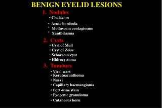
Benign Positional Vertigo Symptoms: Episodes provoked by rolling over in bed, bending over, looking upward Mechanism: debris within posterior canal Proceeded by head trauma or vestibular neuritis Diagnostics: positive dix hallpike. Negative neuro exam. Treatment: Epley maneuver
Who is safe to send home without further evaluation: • Respond well to Epley • Normal neurologic exam • Classic positive Dix Hallpike test: same symptoms are provoked by the maneuver but may or may not provoke the nystagmus
But the Dix Hallpike has a positive predictive value of 83 percent and a negative predictive value of 52 percent for the diagnosis of BPPV.
Case 2: 70yom hx BPH p/w acute vertigo. HPI: While driving, suddenly sensation of being on a boat. Started about 4 hours ago. Unable to walk. N/V x2 Exam: right beating nystagmus on rightward gaze less so on leftward gaze, face symmetric, hearing intact. No worsening of vertigo or nystagmus on Dix Hallpike. Head thrust with catch up saccades. Agrees to stand but gets very angry with you. Romberg falling toward the left twice. Diagnostics: CT shows global atrophy
Clinical exam findings to distinguish peripheral vs. central lesions causing vertigo • Type of nystagmus • Severity of postural stability • Presence of any additional neurologic signs Hotson, J. Acute Vestibular Syndrome. New England Journal. 1998
Inclusion criteria: Acute onset vertigo N/V/retching Gait instability One or more stroke RF Nystagmus Results: Neg HIT c/w incipient lateral medullary stroke (iMRI nl) Pos HIT: missed some, next 24hr neuro exams developed my sxs
Prevalence of dizziness in ER’s: 7.5million patients/year in USA • Prevalence of stroke in patients with acute vestibular syndrome estimated 25% • 16% AICA strokes can present with a peripheral presentation with unidirectional nystagmus • 42% of strokes causing an acute vestibular syndrome do NOT have obvious central findings • Head impulse test can also be positive in a central etiology
Work Up When to immediately obtain brain imaging on a pt with vertigo: • Hx possible for cerebellar hemorrhage • Exam not COMPLETELY c/w peripheral vestibular process • Sudden onset of sxs with stroke risk factors • New severe HA along with vertigo
Work Up When imaging can be delayed 48hours: • Isolated acute vertigo • Peripheral nystagmus suppressed by visual fixation, unstable but can still walk • Lots improvement in 48hrs (vestibular neuritis, no need to image) • Concurrent acute hearing loss, strongly peripheral
Work Up Type of imaging: MRI/MRA. Also gadolinium with fine cuts through CN8 and cerebellopontine angle MRI/MRA within 48hrs with neuro monitoring in between.