Download
1 / 60
640 likes | 814 Views
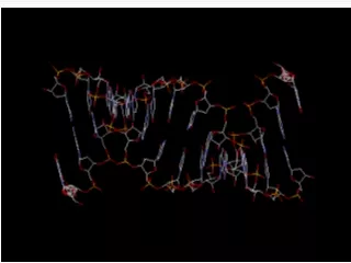
Human Genetics. (Chapter 3: and some of 4). The structure of DNA. Composed of 4 nucleotide bases, 5 carbon sugar and phosphate. Base pair = rungs of a ladder. Edges = sugar-phosphate backbone. Double Helix Anti-Parallel . Figure 2. 21. The structure of DNA. Figure 2.22a.

E N D
Human Genetics (Chapter 3: and some of 4)
The structure of DNA • Composed of 4 nucleotide bases, 5 carbon sugar and phosphate. • Base pair = rungs of a ladder. • Edges = sugar-phosphate backbone. • Double Helix • Anti-Parallel
Figure 2.21 The structure of DNA
Figure 2.22a DNA Replication Remember – the two strands run in opposite directions Synthesis of a new (daughter) strand occurs in the opposite direction of the old (parental) strand. Complementary base-pairing occurs A with T and G with C G and C have three hydrogen bonds A and T have two hydrogen bonds
DNA Replication • Each new double helix is composed of an old (parental) strand and a new (daughter) strand. • As each strand acts as a template, process is called Semi-conservative Replication. • Replication errors can occur. Cell has repair enzymes that usually fix problem. An error that persists is a mutation. • This is permanent, and alters the phenotype.
The structure of RNA • Formed from 4 nucleotides, 5 carbon sugar, phosphate. • Uracil is used in RNA. • It replaces Thymine • The 5 carbon sugar has an extra oxygen. • RNA is single stranded.
Central Dogma of Molecular Biology • DNA holds the code • DNA makes RNA • RNA makes Protein • DNA to DNA is called REPLICATION • DNA to RNA is called TRANSCRIPTION • RNA to Protein is called TRANSLATION
Central Dogma of Molecular Biology • There are exceptions: • Retroviruses • Use RNA as the genetic code • Must make DNA before making protein product • This new DNA makes RNA and then a protein • Also, one protein is not always the product of a single gene – we will talk about this later in the course!
Figure 3.3 (1) Transcription – DNA to RNA (RNA polymerase)
Figure 3.3 (2) Transcription – DNA to RNA
Figure 3.3 (3) Transcription – DNA to RNA
Figure 3.3 (4) Transcription – DNA to RNA
A close-up view of transcription RNA nucleotides RNA polymerase Newly made RNA Direction of transcription Template strand of DNA
How does the order or sequence of nucleotides in a DNA and then a RNA molecule determine the order of amino acids in a protein? (Translation) TACCTGAACGTACGTTGCATGACT DNA RNA AUGGACUUGCAUCGAACGUACUGA Met-Asp-Leu-His-Arg-Thr-Tyr-STOP protein
Translation • Translation requires: • Amino acids (AAs) • Transfer RNA: (tRNA) Appropriate to its time, transfers AAs to ribosomes. The AA’s join in cytoplasm to form proteins. 20 types. Loop structure • Ribosomal RNA: (rRNA) Joins with proteins made in cytoplasm to form the subunits of ribosomes. Linear molecule. • Messenger RNA: (mRNA) Carries genetic material from DNA to ribosomes in cytoplasm. Linear molecule.
Translation • The mRNA has aspecific “open reading frame” made up of three base pairs –codon. • The tRNA has the complementary base-pairing fit to the codon –known as an Anticodon • Each of these codes for an amino acid
Translation • Initiation— • mRNA binds to smaller of ribosome subunits, then, small subunit binds to big subunit. • AUG start codon--complex assembles • Elongation— • add AAs one at a time to form chain. • Incoming tRNA receives AA’s from outgoing tRNA. Ribosome moves to allow this to continue • Termintion— Stop codon--complex falls apart
Figure 3.5 (1) Translation
Figure 3.5 (2) Translation
Figure 3.5 (3) Translation
What happens when it all goes wrong? • MUTATIONS!!!!!!!!!! • two general categories 1.result in changes in the amino acids in proteins A change in the genetic code 2.Change the reading frame of the genetic message Insertions or deletions
Figure 3.6a Mutations
Remember Thalidomide? • The structure of thalidomide is similar to that of the DNA purine bases adenine (A) and guanine (G). • In solution, thalidomide binds more readily to guanine than to adenine, and has almost no affinity for the other nucleotides, cytosine (C) and thymine (T). • Furthermore, thalidomide can intercalate into DNA, presumably at G-rich sites.
Remember Thalidomide? • Thalidomide or one of its metabolites intercalates into these G-rich promoter regions, inhibiting the production of proteins and blocking development of the limb buds. • This intercalation would not significantly affect the over 90 per cent of genes that rely primarily on guanine sequences. • Most other developing tissues in the embryo rely on pathways without guanine, and are therefore not affected by thalidomide
Genes can lead to inherited diseases • A gene which doesn’t function on an autosomal chromosome can lead to devastating diseases • Autosomal chromosomes are 22 pairs of chromosomes which do not determine gender • Such diseases can be caused by both a dominant or a recessive trait
Autosomal Recessive Disorders • Tay-Sachs Disease: • Jewish people in USA (E. Euro descent) • Not apparent at birth • 4 to 8 months • Neurological impairment evident • Gradually becomes blind and helpless • Develops uncontrollable seizures/paralyzed • Allele is on Chromosome 15 • Lack of enzyme hexosaminidase A (Hex A) • Lysosomes don’t work, build up in brain
Autosomal Recessive Disorders • Cystic Fibrosis • Most commonin USA(Caucasian) • 1 in 20 caucasians is a carrier • Mucus in bronchial and pancreas thick/viscous • Breathing and food digestion problems • Allele is on chromosome 7 • Cl ions can not pass through plasma membrane channels • Cl ions pass –water goes with it. No water, thick mucus
Autosomal Recessive Disorders • Phenylketonuria (PKU) • Affects in in 5,000 newborns • Most common nervous system disorder • Allele is on chromosome 12 • Lack the enzyme needed for the metabolism of the amino acid phenylalanine • A build up of abnormal breakdown pathway • Phenylketone • Accumulates in urine. If diet is not checked, can lead to severe mental retardation
Autosomal Dominant Disorders • Neurofibromatosis • Very common genetic disorder • Tan spots on skin • Later tumors develop • some sufferers have large head and ear and eye tumors. • Allele is on chromosome 17 • Gene controls the production of a protein called neurofibromin • This naturally stops cell growth
Autosomal Dominant Disorders • Huntington Disease • Leads to degeneration of brain cells • Severe muscle spasms and personality disorders • Attacks in middle age • Allele is on chromosome 4 • Gene controls the production of a protein called huntington • Too much AA glutamine. Changes size and shape of neurons
Incomplete Dominant traits • Sickle Cell Anemia • Controlled by intermediate phenotypes at a ratio of 1:2:1 • Red blood cells are not concave • Normal Hemoglobin (HbA). Sickle cell (HbS) • HbA-HbA-normal Hbs-Hbs – sickle cell • HbA-Hbs- have the trait
Mutations - any change in the nucleotide sequence of DNA Normal hemoglobin DNA Mutant hemoglobin DNA mRNA mRNA Sickle-cell hemoglobin Normal hemoglobin Glu Val Figure 10.21
Individual homozygousfor sickle-cell allele Sickle-cell (abnormal) hemoglobin Abnormal hemoglobin crystallizes, causing red blood cells to become sickle-shaped Sickled cells Clumping of cells and clogging of small blood vessels Breakdown of red blood cells Accumulation of sickled cells in spleen Damage to other organs Pain and fever Brain damage Heart failure Physical weakness Spleen damage Anemia Impaired mental function Pneumonia and other infections Kidney failure Rheumatism Paralysis Figure 9.21
Genetic engineering • The direct alteration of a genotype • Human genes can be inserted into human cells for therapeutic purposes • Genes can be moved from one species to another • Moving genes from human to human or between species requires the use of special enzymes known as restriction enzymes. • These cut DNA at very specific sites • They restrict DNA from another species – isolated from bacteria.
Figure 4.1 Genetic engineering • Each restriction enzyme cuts the DNA at a specific site, defined by the DNA sequence • Enzymes which produce “sticky ends” are more useful • Allows gene of interest to be inserted into a vector • Also need a DNA probe • Radioactive ssDNA that will bind to gene of interest so you can locate it
Genetic engineering • Transferred DNA is denatured to give ssDNA • The probe will bind to gene of interest by Complementary base-pairing - A with T and G with C
Figure 4.3 (1) Genetically engineered insulin
Figure 4.3 (2) Genetically engineered insulin
Figure 4.3 (3) Genetically engineered insulin
Figure 4.3 (4) Genetically engineered insulin
Genetically engineered insulin • Why do some people not like the idea? The plasmid also needs a “marker gene” This is usually an antibiotic resistance gene Some people fear that the insulin which is extracted from the bacteria would also contain a gene product to make anyone who uses the insulin resistant to antibiotics!
Gene therapy • Can treat human diseases • eg – severe combined immune deficiency syndrome (SCIDS) • Bubble- Boy/Girl syndrome • The enzyme which causes this is on chromosome 20 • Called adenosine deaminase(ADA) • Many problems • Difficult to transfer large genes • Insert in a way that the gene expresses to protein correctly • TRANSLATION!!!!!!!!!!!!
Figure 4.4 (1) Gene therapy