Download
1 / 47
620 likes | 1.53k Views
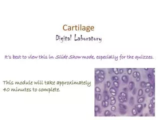
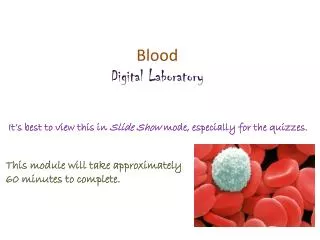
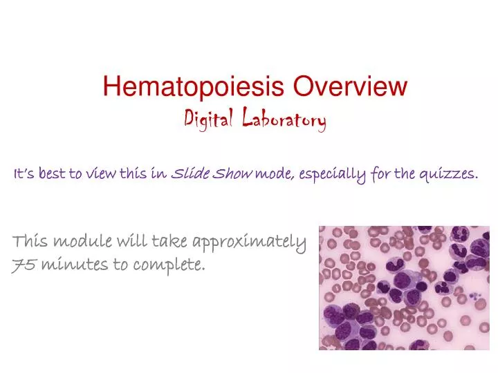
Hematopoiesis Overview Digital Laboratory. It’s best to view this in Slide Show mode, especially for the quizzes. This module will take approximately 75 minutes to complete. After completing this exercise, you should be able to:

E N D
Hematopoiesis Overview Digital Laboratory It’s best to view this in Slide Show mode, especially for the quizzes. This module will take approximately 75 minutes to complete.
After completing this exercise, you should be able to: • identify, at the light microscope level, each of the following: • Cytoplasmic granules • Azurophilic(nonspecific) granules • Basophilic granules • Eosinophilic granules • Neutrophilicgranules • Nucleoli (warning, these ain’t your father’s nucleoli) • Nuclear shape • Round • Indented • Kidneybean-shaped • Band • Segmented • Chromatin pattern • Fine • Clumped • Cytoplasmic staining • Cytoplasmic basophilia • Cytoplasmic eosinophilia
Five cytological features are used to differentiate amongst developing blood cell types. They are: cytoplasmic granules presence or absence of nucleoli shape of the nucleus extent of nuclear chromatin clumping degree of cytoplasmic basophilia Note that blood cells are stained routinely with specific hematologic dyes, not H&E, and this results in a slightly wider range of coloration than seen with H&E. A new color is recognized - azure, a bluish purple. To view critical cytologic features of blood cells, images in your hematology lab modules are magnified to a much greater degree than seen in previous labs. To gauge relative magnification, red blood cells (7.5 µm in diameter) in the field of view are a good yardstick.
CYTOPLASMIC GRANULES azurophilic (primary, or nonspecific) granulesare purple and readily visualized because of their size. These cells have many; some are indicated at the arrows. The azurophilic granules are described as nonspecific because their appearance during granulocyte maturation is no help in distinguishing amongst precursors of the neutrophil, eosinophil, and basophil lineage. The other three granules (on the following slides) are called specific because their appearance allows one to definitively place the cell in a specific granulocyte lineage.
CYTOPLASMIC GRANULES eosinophilic granulesare very large, red-pink, and have a glassy appearance. The two cells within the black circles are chock-full of these granules.
CYTOPLASMIC GRANULES basophilic granulesare very large and blue. In this mature basophil, note the large granules indicated by the black arrows.
CYTOPLASMIC GRANULES neutrophilic granulesare too small (0.1-0.2 um) to be seen as individual structures with the light microscope. Their presence in neutrophilic granulocytes is signified by a salmon-colored appearance of the cytoplasm. As Cell A, a granulocyte precursor with a basophilic cytoplasm and many azurophilicgranules, matures to stage B, then C, then D, synthesis of neutrophilic granules renders the cytoplasm pink. C B C D A
CYTOPLASMIC GRANULES Video of nonspecific (azurophilic, primary) granules – SL68 Video eosinophilic granules – SL177 Video basophilic granules – SL177 Video neutrophilic granules – SL177 • Link to SL 068and SL 177 • Be able to identify: • Azurophilic (nonspecific, primary) granules • Specific granules • Eosinophilic granules • Basophilic granules • Neutrophilic granules (recognize the presence of these)
NUCLEOLI presence or absence of nucleoli – Cells in the earliest recognizable stages of blood cell development have nucleoli. With hematologic stains, these nucleoli appear as palespots in a darker nuclear background; 1-3/nucleus may be apparent. Note that with histological stains you are familiar with thus far, nucleoli were seen as dark densities within a pale, euchromatic nucleus….the opposite is true here. Advance slide to see arrows on nucleoli…I suggest toggling back and forth a few times. Note: not all possible nucleoli are indicated.
NUCLEOLI Video of nucleoli – SL177 • Link to SL 177 • Be able to identify: • nucleoli
NUCLEAR SHAPE In the early stages of all lineages of developing white blood cells, nuclei tend to be large and round. In the early stages below, the nuclear envelope is indicated by the tips of the red arrows. You may observe some irregularities caused by impinging adjacent cells and/or tissue preparation. Advance slide to see arrows
NUCLEAR SHAPE All recognizable cells in the red blood cell (RBC) lineage maintain a round shaped-nucleus during maturation, though the nuclei become significantly smaller. For example, the cell marked A is the earliest RBC precursor, and the cell marked D is the latest nucleated RBC precursor. B and C are intermediates. Again, the nuclear envelope is indicated by the tips of the arrows. D C B D A
NUCLEAR SHAPE Cells of the granulocyte lineages are quite different. During maturation, the early round nucleus (A) undergoes shape changes that include indentations (B) and then seg-mentation (C). This is easiest to see in neutrophils because nuclei are readily visible. B C B B B A
NUCLEAR SHAPE In eosinophils and basophils, nuclei are less prominently segmented. In addition, the cytoplasmic granules often make it difficult to distinguish the exact shape of the nucleus. Nevertheless, these maturing forms do demonstrate nuclear indentation, as in the eosinophil to the right, and complete segmentation, as demonstrated by the basophil to the left. eosinophil neutrophil basophil
NUCLEAR SHAPE Video of nuclear shape – SL177 • Link to SL 177 • Be able to identify: • Appreciate changes in nuclear shape • Round • Indented • Kidney-bean • Band • Segmented
CHROMATIN The chromatin content of the nucleus is described as either fine (thread-like), or clumped. In the early stages of blood cell development shown here (when nucleoli are visible) the chromatin in the remainder of the nucleus is fine. In the circled cell on the right, a few azurophil granules overlie the nucleus. These must be mentally subtracted in assessing the chromatin pattern.
CHROMATIN As blood cells mature, chromatin often becomes clumped. A good example of this is shown in the basophilic erythroblast (red arrow)….the clumping of the chromatin in this nucleus results in a distinctive dark, ropy pattern (solid black arrows) that leaves some areas white (dashed black arrows). An earlier stage of erythroblast is shown to the left for comparison. A similar dark and ropy staining pattern due to condensation of chromatin also is easily seen in the nuclei of developing granulocytes (blue arrows). An extreme kind of condensation called pyknosis is seen in late stage erythroblasts (green arrows), in which the chromatin condenses completely and the nucleus is reduced in size.
CHROMATIN Video of chromatin – SL177 • Link to SL 177 • Be able to identify: • Fibrillar chromatin • Clumpy chromatin
DEGREE of CYTOPLASMIC BASOPHILIA
CYTOPLASMIC BASOPHILIA Generally speaking, immature cells have a basophilic cytoplasm due to large amounts of ribosomal RNA. In the case of erythroblasts preparing to make hemoglobin, this is in the form of free ribosomes. In granulocytes, the basophilia is due to rough ER. In these early cells outlined in blue, you can see the intense cytoplasmic basophilia.
CYTOPLASMIC BASOPHILIA In general, as cells mature, there is a gradual decrease in the rRNA content and a concomitant increase of protein or organelles that render the cytoplasm less basophilic and more eosinophilic. In neutrophil development, the cytoplasm gets pinker due to accumulation of neutrophil granules. The cells on the left (pink asterisks) are in an early stage of development and have a roughly evenly blue cytoplasm. The two circled cells to the right have accumulated granules in the region of the Golgi, resulting in a prominent patch of pink (pink arrows) in the formerly exclusively blue cytoplasm. Gradually, the cytoplasm of the mature neutrophil appears to become exclusively pink (black arrows). * *
CYTOPLASMIC BASOPHILIA In the developing RBC lineage, the rRNA and hemoglobin are cytosolic. Therefore, the mix of pink/blue color possibilities are greater than in the developing neutrophils. The erythrocyte cytoplasm is pure blue (red arrow) at early stages; as the cells mature (green arrow), eosinophilic hemoglobin is added, resulting in a dull purple or grey color. As the rRNA is lost in more mature cells, the cytoplasm becomes progressively pinker (blue arrow). 1 3 2
CYTOPLASMIC BASOPHILIA Video of cytoplasm color change in the neutrophic series – SL177 Video of cytoplasm color change in the erythrocyte series part 1 – SL68 Video of cytoplasm color change in the erythrocyte series part 2 – SL177 • Link to SL 177and SL 068 • Be able to identify: • Maturity of cell based on cytoplasmic basophilia/eosinophilia
The next set of slides is a quiz for this module. You should review the structures covered in this module, and try to visualize each of these in light. • identify, at the light microscope level, each of the following: • Cytoplasmic granules • Azurophilic(nonspecific) granules • Basophilic granules • Eosinophilic granules • Neutrophilicgranules • Nucleoli (warning, these ain’t your father’s nucleoli) • Nuclear shape • Round • Indented • Kindey-bean-shaped • Band • Segmented • Chromatin pattern • Fine • Clumped • Cytoplasmic staining • Cytoplasmic basophilia • Cytoplasmic eosinophilia
Final quiz Self-check: Does this cell have nucleoli? If so, how many nucleoli do you see?? Yes 3 (though you may have seen more or less)
Final quiz Self-check: Identify the structures at the tips of the arrows eosinophilicgranules
Final quiz Self-check: Does the outlined cell have cytoplasmic granules? If so, what type? Yes, it has a ton of nonspecific (azurophilic, primary) granules
Final quiz Self-check: On a scale of 1 to 10, 1 being the least mature, 10 being a red blood cell, where would you place this cell based on its cytoplasmic color? 7 (6-8)
Final quiz Self-check: Does the outlined cell have nucleoli? If so, how many nucleoli do you see?? No
Final quiz Self-check: Does this cell have nucleoli? If so, how many nucleoli do you see?? Yes 2
Final quiz Self-check: What is the term that describes the shape of the nucleus of this cell? band
Final quiz Self-check: Does this cell have nucleoli? If so, how many nucleoli do you see?? No
Final quiz Self-check: Identify (yes, name) the outlined cell? lymphocyte
Final quiz Self-check: Identify the structures at the tips of the arrows Nonspecific (azurophilic, primary) granules
Final quiz Self-check: Of cell A or B, which has the finer chromatin pattern? Which has the nucleolus? B A B
Final quiz Self-check: Identify the structures at the tips of the arrows. eosinophilic granules
Final quiz Self-check: Identify the structures at the tips of the arrows. Basophilic granules
Final quiz Self-check: On a scale of 1 to 10, 1 being the least mature, 10 being a red blood cell, where would you place this cell based on its cytoplasmic color? 1 (or 2)
Final quiz Self-check: Place these three cells (A-C) in the neutrophil series in order based on increasing maturity. A B B, A, C C
Final quiz Self-check: Identify the structures at the tips of the arrows eosinophilicgranules Compare these to the granules in the WBC that is cut off at the top. Also, the “dude” in the bottom right looks soooocoooool in those shades.
Final quiz Self-check: On a scale of 1 to 10, 1 being the least mature, 10 being a red blood cell, where would you place this cell based on its cytoplasmic color? 5 or 6
Final quiz Self-check: Identify (yes, name) the outlined cell? This is for super-double bonus points. Plasma cell
OK, I’ve squeezed out about as much as I could from this module. For the weekly quiz, expect some quiz questions similar to these. However, the ultimate goal is for you to identify specific cell types, which will be the focus of subsequent modules.