Download
1 / 17
180 likes | 554 Views
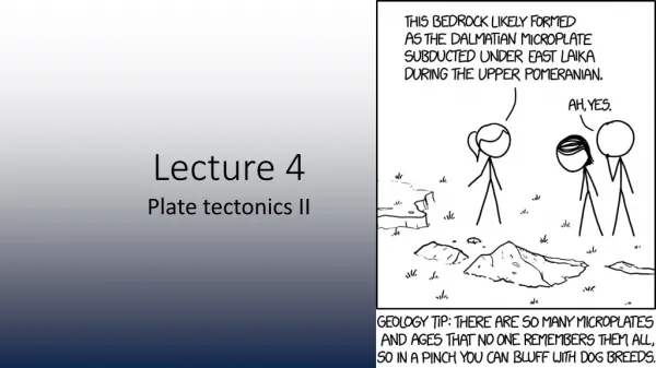
Lecture 4. 13 C NMR: DEPT IR Spectroscopy: - How it works Interpretation of spectra Due: Lecture Problem 2. Determine the structure of this unknown (MF is C 8 H 9 Cl). 13 C NMR Correlation Chart. 1 H NMR Correlation Chart. DEPT-NMR

E N D
Lecture 4 • 13C NMR: DEPT • IR Spectroscopy: • - How it works • Interpretation of spectra • Due: • Lecture Problem 2
13C NMR Correlation Chart 1H NMR Correlation Chart
DEPT-NMR (Distortionless Enhancement by Polarization Transfer) • Distinguishes between CH, CH2, and CH3 carbons
13C NMR: broadband decoupled (normal) 13C NMR: DEPT-90 13C NMR: DEPT-135
MRI: A Medicinal Application of NMR • Magnetic Resonance Imaging: • MRI Scanner: large magnet; coils to excite • nuclei, modify magnetic field, and receive • Signals • Different tissues yield different signals • Signals are separated into components by • Fourier transform analysis • Each component is a specific site of origin • in the patient a cross-sectional image of • the patient’s body • How it works: • Most signals originate from hydrogens of • Water molecules • Water is bound to different organs in different way variation of signal among • organs & variation between healthy and diseased tissue MRI showing a vertical Cross section through a Human head. http://en.wikipedia.org/wiki/Magnetic_resonance_imaging
MRI: A Medicinal Application of NMR • Some Magnetic Resonance Imaging Uses: • Detailed images of blood vessels • Examine the vascular tree • Differentiate intracelluar and • extracelluar edema stroke patients • Detecting cancer, inflammation, tumors • Current research: • 31P nuclei analysis: investigate celluar • metabolism (ATP and ADP) MRI showing a vertical Cross section through a Human head. http://en.wikipedia.org/wiki/Magnetic_resonance_imaging
Spectroscopy 1H NMR: Determine bond connectivities/pieces of a structure, whole structure 13C NMR: Types of carbons (DEPT) IR: Determine the functional groups present in a structure: -OH, C=O, C-O, NH2, C=C, CC, C=N, CN
IR Spectroscopy Main Use: To detect the presence or absence of a functional group (specific bonds) in a molecule How It Works: Bonds vibrate freely at specific wavelengths (wavenumbers) Want to cause the bonds to increase the magnitude of this vibrational frequency Subject compound to IR radiation, 4000-625 cm-1 cm-1 is the unit for wavenumber (n) n is directly proportional to energy (unlike wavelength) 4. Bonds absorb energy equal to their natural vibrational energy - it is quantized. This absorption of energy causes a change in dipole moment for the bond. 5. Upon absorption, bonds stretch and/or bend; the IR measures this absorption.
Vibrational Modes of Bonds Stretches are more noted than bends
Correlation Chart Specific bonds absorb specific IR radiation and signals will appear within certain wavenumber ranges (similar to NMR). Note: O-H stretches are broader than N-H stretches N-H Stretches: 1° Amines (RNH2) has two peaks 2° Amines (RNHR) has one peak 3° Amines (NR3) has no peaks
IR Correlation Chart Specific bonds absorb specific IR radiation and signals will appear within certain wavenumber ranges (similar to NMR).
A: O-H stretch (strong, broad) C: C-H stretch (strong, sharp) E: CC or CN stretch (sharp) F: C=O stretch (strong, medium to sharp) G: C=C stretch (sharp) J: C-O stretch (strong, medium) K: C-X stretch (sharp)
IR spectrum of hexanoic acid Fingerprint Region: 400-1550 cm-1 More difficult to interpret Functional Group Region: 1550-4000 cm-1 Most useful portion
An IR Spectrum O-H stretches are broad due to H-bonding.
Sample Problem 1 Indicate how the following pairs of compounds could be distinguished using characteristic IR peaks: (a) Benzaldehyde (C6H5O) and benzoic acid (C6H5COOH) 1. Consider each structure: benzaldehyde Benzoic acid 2. Determine the main differences that would be seen in IR. Use correlation chart.
Sample Problem 2 An unknown oxygen-containing compound is suspected of being an alcohol, a ketone, or a carboxylic acid. Its IR spectrum shows a broad strong peak at 3100-3400 cm-1 and a strong, sharp peak at 1700 cm-1. What kind of compound is it? Consider what type of bonds appear in the ranges given. Refer to correlation chart. Broad peak at 3100-3400 cm-1 Strong, sharp peak at 1700 cm-1