Download
1 / 56
570 likes | 788 Views
PROTEIN SYNTHESIS. Bashdar M. Hussen MSc.Biotechnology Hawler Medical University. Discovering the structure of DNA. 1. DNA was dicovered by Juhann Friedrich in 1869 2. And the first demonstration that DNA contain genetic information was made in 1944 by Avery, Macleod and MacCary

E N D
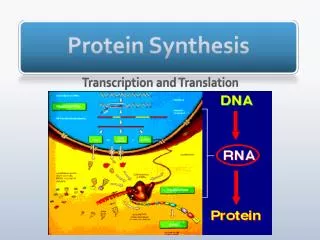
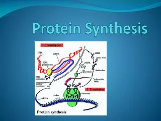
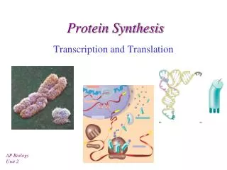
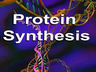
PROTEIN SYNTHESIS BashdarM. Hussen MSc.Biotechnology Hawler Medical University
Discovering the structure of DNA 1. DNA was dicovered by Juhann Friedrich in 1869 2. And the first demonstration that DNA contain genetic information was made in 1944 by Avery, Macleod and MacCary 3. 1951—Rosalind Franklin was showed or explained the —X-ray crystallography of DNA 4. 1953 Watson & Crick—explained completstru. Of DNA
Watson & Crick Model DNAStructure was discovered in 1953 by James Watson and Francis Crick
DNA Structure • A molecule of DNA is made up of millions of tiny subunits called Nucleotides. • Each nucleotide consists of: • Phosphate group • Pentose sugar • Nitrogenous base
DNA is composed of 2 chains of nucleotides that form a double helix shape. The two strands are antiparallel. The backbone of the DNA molecule is composed of alternating phosphate groups and sugars. The complimentary nitrogenous bases form hydrogen bonds between the strands. A is complimentary to T and G is complimentary to C. The Diameter is 20 A(2nm) The 2 polynucleotide chains are not identical but complimentary to each other. The hydrogen bonds formed between purine and pyrimidine only. The DNA is write handed double helix.
Synthetic Nucleotide analogs & Chemotherapy Synthetic analogs of purines, pyrimidines, nucleosides, and nucleotides altered in either the heterocyclic ring or the sugar moiety. 1. The purine analog allopurinol, used in treatment of hyperuricemia and gout. 2. Cytarabine is used in chemotherapy of cancer. 3. Azathioprine is employed during organ transplantation to suppress immunologic rejection.
phenotype DNA Protein Gene Gene Trait
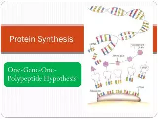
Genes & Proteins • DNA contains genes, sequences of nucleotide bases • These Genes code for polypeptides (proteins) • Proteins are used to build cells and do much of the work inside cells • Proteins are made of amino acids linked together by peptide bonds • 20 different amino acids exist
DNA Begins the Process • DNA is found inside the nucleus • Proteins, however, are made in the cytoplasm of cells by organelles called ribosomes • Ribosomes may be free in the cytosol or attached to the surface of rough ER
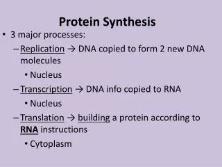
Starting with DNA • DNA ‘s code must be copied and taken to the cytosol • In the cytoplasm, this code must be read so amino acids can be assembled to make polypeptides (proteins) • This process is called PROTEIN SYNTHESIS
Roles of RNA and DNA • DNA is the MASTER PLAN • RNA is the BLUEPRINT of the Master Plan
Other Differences • RNA contains the base uracil (U) DNA has thymine (T) • RNA molecule is single-stranded DNA is double-stranded DNA
Three Types of RNA . • Messenger RNA (mRNA) copies DNA’s code & carries the genetic information to the ribosomes • Ribosomal RNA (rRNA), along with protein, makes up the ribosomes • Transfer RNA (tRNA) transfers amino acids to the ribosomes where proteins are synthesized
Messenger RNA • Long Straight chain of Nucleotides • Made in the Nucleus • Copies DNA & leaves through nuclear pores • Carries the information for a specific protein • Made up of 500 to 1000 nucleotides long • Sequence of 3 bases called codon • AUG – methionine or start codon • UAA, UAG, or UGA – stop codons
Ribosomal RNA (rRNA) • rRNA is a single strand 100 to 3000 nucleotides long • Globular in shape • Made inside the nucleus of a cell • Associates with proteins to form ribosomes • Site of protein Synthesis
The Genetic Code • A codon designates an amino acid • An amino acid may have more than one codon • There are 20 amino acids, but 64 possible codons • Some codons tell the ribosome to stop translating
Name the Amino Acids • AG? • UCA?
Remember the Complementary Bases On DNA: A-T C-G On RNA: A-U C-G
Transfer RNA (tRNA) • Clover-leaf shape • Single stranded molecule with attachment site at one end for an amino acid • Opposite end has three nucleotide bases called the anticodon
Codons and Anticodons • The 3 bases of an anticodon are complementary to the 3 bases of a codon • Example: Codon ACU Anticodon UGA UGA ACU
Transcription • The process of copying the sequence of one strand of DNA, the template strand • mRNA copies the template strand • Requires the enzyme RNA Polymerase
Question: • What would be the complementary RNA strand for the following DNA sequence? DNA 5’-GCGTATG-3’
Transcription • Consists of three stages • Initiation: attachment of RNA Polymerase to the promotor region on DNA • Elongation: building of the mRNA from the 3’ end of the nucleotide polymer • Termination: release of RNA polymerase and mRNA following transcription of the terminator region of the DNA
1. Initiation • Genes on the DNA begin with a promoter region consisting of a sequence of A & T (TATA box) • Transcription factors (proteins that assist the binding of RNA polymerase to the promoter) are found in association with the promoter region
Elongation • Once initiation is complete the 2 strands of the DNA unwind due to the zipper region of the enzyme. • RNA polymerase builds a mRNA strand complimentary to the DNA transcription unit. • Once the RNA Polymerase passes the DNA strands reform their double helix
Termination In Eukaryotes • The transcribed termination sequence, also known as the polyadenylation signal in the pre-mRNA, is AAUAAA. • Polymerase continues to synthesize RNA until an enzyme catches up to it and causes it too fall off.
Modification of mRNA • Transcribed mRNA (pre-mRNA) must be modified before leaving the nucleus • modifications include: • addition guanine triphosphate cap to the 5” end of the mRNA • Prevents “unraveling” • Helps ribosome attach • addition of poly A tail to the 3’ end of the mRNA • Prevents “unraveling” • Assists in the export of mRNA from nucleus
Result of Transcription New Transcript Tail CAP
How is this done? • Small nuclear ribonucleicproteins (snRNP) recognize intron ends and together with proteins form a structure called a spliceosome • Spliceosomes remove introns while connecting exons together
Why bother with introns? • Introns may regulate gene activity and the passage of mRNA into the cytoplasm • Genes may play roles in multiple proteins, introns may enable a gene to be diverse in function • May increase recombination of genetic material (easier to cut and paste)
Translation • Translation-forming of a polypeptide -uses mRNA as a template for a.a. sequence -steps (initiation, elongation, translocation and termination) -begins after mRNA enters cytoplasm -uses tRNA (the interpreter of mRNA)
Translation B. Ribosomes -made of proteins and rRNA -each has a large and small subunit -each has three or two binding sites for tRNA on its surface -each has one binding site for mRNA -facilitates codon and anticodon bonding
Translation -The three tRNA binding sites are: 1. A site=holds tRNA that is carrying the next amino acid to be added 2. P site= holds tRNA that is carrying the growing polypeptide chain 3. E site= where discharged tRNAs leave the ribosome
Translation • 61 of 64 codons code for a.a. • Codon AUG has two functions 1. codes for amino acid methionine (Met) 2. functions as a start codon • mRNA codon AUG starts translation • three codons act as stop codons (end translation)
A. (Initiation) 1. Brings together mRNA, tRNA (w/ 1sta.a.) and ribosomal subunits 2. Small ribosomal subunit binds to mRNA and an initiator tRNA -start codon= AUG - anticodon-UAC -small ribosomal subunit attaches to 5’ end of mRNA
-downstream from the 5’ end is the start codon AUG (mRNA) -the anticodon UAC carries the a.a. Methionine 3. After the union of mRNA, tRNA and small subunit, the large ribosomal subunit attaches. • The intitiatortRNA and a.a. will sit in the P site of the large ribosomal subunit • The A site will remain vacant and ready for the aminoacyl-tRNA
B. (Elongation) Amino acids are added one by one to the first amino acid (remember, the goal is to make a polypeptide) Step 1: Codon recognition mRNA codon in the A site forms hydrogen bonds with the tRNAanitcodon Step 2- Peptide bond formation The ribosome catalyzes the formation of the peptide bonds between the amino acids (the one already in place and the one being added) The polypeptide extending from the P site moves to the A site to attach to the new amino acid.
Translation (Translocation) • The tRNA with the polypeptide chain in the A site is translocated to the P site • tRNA at the P site moves to the E site and leaves the ribosome • The ribosome moves down the mRNA in the 5’→3’ direction
aa2 aa1 2-tRNA 1-tRNA G A U U A C Initiation anticodon A U G C U A C U U C G A hydrogen bonds codon mRNA
aa3 3-tRNA G A A Elongation peptide bond aa1 aa2 1-tRNA 2-tRNA anticodon U A C G A U A U G C U A C U U C G A hydrogen bonds codon mRNA
aa3 3-tRNA G A A aa1 peptide bond aa2 1-tRNA U A C (leaves) 2-tRNA G A U A U G C U A C U U C G A mRNA Ribosomes move over one codon
aa4 4-tRNA G C U peptide bonds aa1 aa2 aa3 2-tRNA 3-tRNA G A U G A A A U G C U A C U U C G A A C U mRNA
aa4 4-tRNA G C U peptide bonds aa1 aa2 aa3 2-tRNA G A U (leaves) 3-tRNA G A A A U G C U A C U U C G A A C U mRNA Ribosomes move over one codon