Download
1 / 14
140 likes | 230 Views
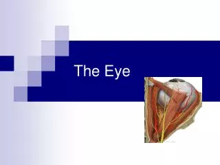
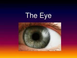
The Eye. The Physiology of Vision. Anatomy of the Eye. The human eye is a complex organ that is sensitive to a very specific range of electromagnetic radiation

E N D
The Eye The Physiology of Vision
The human eye is a complex organ that is sensitive to a very specific range of electromagnetic radiation • The eye allows us to see and interpret the shapes, colors, and dimensions of objects in the world by converting the light waves they reflect or emit into nerve signals. • The eye is able to detect bright light, dim light and colour, but it cannot sense objects when light is absent. (no light = no nerve impulses)
The Process of Vision Light waves from an object enter the eye first through the cornea. The light then progresses through the pupil, the circular opening in the center of the colored iris.
Fluctuations in the intensity of incoming light change the size of the eye’s pupil. As the light entering the eye becomes brighter, the pupil will constrict (get smaller), due to the pupillary light response. As the entering light becomes dimmer, the pupil will dilate (get larger).
Initially, the light waves are bent or converged first by the cornea, and then further by the crystalline lens (located immediately behind the iris and the pupil), to a point located immediately behind the back surface of the lens. At that point, the image becomes reversed (turned backwards) and inverted (turned upside-down).
The light continues through the vitreous humor, the clear gel that makes up about 80% of the eye’s volume, and then back to a clear focus on the retina, behind the vitreous. The small central area of the retina is the macula, which provides the best vision of any location in the retina.
Within the layers of the retina, light impulses are changed into electrical signals. Then they are sent through the optic nerve to the occipital cortex at the posterior (back) of the brain. Here, the electrical signals are interpreted or “seen” by the brain as a visual image.