Download
1 / 44
830 likes | 3.04k Views
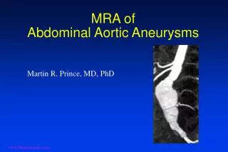
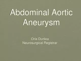
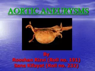
Abdominal Aortic Aneurysms. Aurelia Thibonnier-Calero PGY-2 Vascular Surgery. Types of Aneurysms. True vs. False (pseudoaneurysm) True : involves all 3 layers of the arterial wall

E N D
Abdominal Aortic Aneurysms Aurelia Thibonnier-Calero PGY-2 Vascular Surgery
Types of Aneurysms • True vs. False (pseudoaneurysm) • True: involves all 3 layers of the arterial wall • False: presence of blood flow outside of normal layers of arterial wall. Wall of false aneurysm is compose of the compressed, surrounding tissues.
Types of Aneurysms • Etiology • Degenerative- complex process that involves some degree of calcification, atherosclerotic pathology as well as degeneration by MMPs. • Inflammatory- thick inflammatory wall with fibrotic process in retroperitoneum that can encase aorta as well as surrounding structures. Associated with other inflammatory conditions : Takayasu’s, Giant cell arteritis, Polyarteritis nodosa, Behcet’s, Cogans’. • Post-dissection- up to 20% of aneurysms are related to previous dissection. Overtime, develops into true aneurysm • Traumatic- false aneurysms • Developmental Anomalies- persistent sciatic arteries, aberrant right subclavian artery. • Infectious- Can be primary or secondary infections. • Congenital- Tuberous sclerosis, aortic coarctation, Marfan’s.
Assessing the AAA patient • Normal - aorta 1-2.4cm & iliac 0.6-1.2cm • Aneurysm - Aorta >3cm & iliac > 2cm • RF for aneurysm • Older age, male gender, white race, positive family history, smoking, HTN, hypercholesterolemia, PVD, CAD. • Ultrasound • used to diagnose and monitor AAA until aneurysm approaches size at which repair considered. • Computed Tomography • used in preop assessment of AAA.
Ruptured AAA • No significant overall change in mortality with open repair from 1991-2006 • Overall mortality for ruptured AAA = 90% • Mortality rate for patients who arrive at hosptial alive = 40-70% • High postop mortality rate due to MI, renal failure, and multi-organ failure • Ischemia-reperfusion injury, hemorrhagic shock, lower torso ischemia • rEVAR significantly reduces mortality of ruptured AAA patients (31 vs 50%)
Screening for AAA • US Preventive Services Task Force • Men 65-75 yo who have ever smoked • No for or against men 65-75yo who have never smoked • Does not recommend screening for women • Society of Vascular Surgery, Medicare Screening • Men who have smoked at least 100 cigarettes during their life • men and women with a family history of AAA • Only screen patients who are candidates for repair.
Choosing between Surgery & Observation • Risk for AAA rupture without surgery • Operative risk of repair • Patient’s life expectancy • Personal preferance of patient
1. Risk of Rupture • Size matters: • Aneurysm > 5cm 6-16% and > 7cm 33% annual rupture rate • Wall stress analysis • Saccular aneurysm have higher rate of rupture • HTN, COPD, active smoking are independent predictors of rupture • (+) family hx tend to rupture • Expansion rate
2. Operative Risk of Repair • Mortality after: • elective open AAA ~ 5% • EVAR 1% • 6 independent RF’s for mortality Open repair • Creatinine > 1.8, CHF, EKG detected ischemia, Pulmonary dysfunction, older age, female gender. • Cardiac, pulmonary, renal, and GI risks with each proceudre.
3. Patient’s Life Expectancy • Very difficult to assess due to patient’s co-morbidities • Typical 60yo surviving AAA repair has 13year life-expectacy, 70yo has 10year life-expectancy, and 80 yo has 6 year life-expectancy.
4. Personal Preference of Patient • Fear of AAA vs. Fear of surgery • Anecdotal experiences of friends and family • Procedures provided in community by interventional specialists and surgeons.
Medical Management of AAA • Smoking Cessation-Single most important modifiable risk factor • Exercise Therapy- Evidence suggests may benefit small aneurysms • Beta Blockers- May decrease the rate of expansion? Important cardiovascular effects thus use advocated. • ACE inhibitors- Evidence is mixed, however, implicated in less aneurysm rupture. • Doxycycline • Antibiotic activiety against chlamydia species • Suppresses expression of MMP • Statins - associated with reduced aneurysm expansion rates. Decreases MMP-9 in aneurysm wall.
EVAR vs. OPEN • EVAR-1 and DREAM Trials • Randomized AAA > 5.5 cm to EVAR vs. open repair • Lower 30-day mortality for EVAR (1.6% EVAR vs. 4.6% open) • Peripop mortality and severe complications 4.7% EVAR & 9.8% open repair (DREAM) • Similar all-cause mortality at 2 years • Higher rate of secondary interventions in EVAR group • Total cost of Tx & 4 years of f/u is significantly increased for EVAR.
Transabdominal Approach Previous retroperitoneal surgery Ruptured AAA Exposure of mid/distal portions of visceral vessels or R renal artery R internal or external iliac artery Co-existant abdominal pathology Left-sided vena cava Retroperitoneal Approach Mult. Previous intraperitoneal procedures Abd wall stoma, ectopic/ anomaly of kidney Inflammatory aneurysm Proximal aortic access, endarterectomy of viceral/renal arteries needed Obese patients Fewer GI complications Open Repair
Open Repair-Complications • Cardiac • Pulmonary • Renal • Lower Extremity Ischemia • Spinal Cord Ischemia • Incisional Hernia • 14.2% ventral hernia, 9.7% SBO • Graft Infection
Open Repair Complications:Colon Ischemia • Collaterals from SMA, IMA, internal iliac artery, and profunda femoris supply sigmoid colon • Mortality 40-65%, full-thickness necrosis 80-100% • Occurs in 0.6-3% of elective and 7-27% of ruptured AAA (much more common endoscopically than clinically) • Si/Sx: persistent acidosis & shock, increased WBCs and lactate levels, fluid sequestration, bloody bowel movements. • TX: • Ischemia limited to mucosa/submucosa- npo, IVF, IV abx • Transmural ischemia- bowel resection, fecal diversion, creation of ostomy, washout of abdomen, IV abx.
Open Repair- Concomitant Pathology • Treat the most life-threatening process first • Avoid simultaneous operations that increase the risk for prosthetic graft infection • If secondary procedure can be staged without increased risk - do aneurysm repair first • Clean procedures (ie:nephrectomy, oophrectomy) can be performed simultaneously with open AAA repair • GI procedures should not occur at same time as open repair • Abort surgery if metastatic disease or abscesses which increase risk for graft infection discovered.
Inflammatory AAA • Perianeurysmal fibrosis & inflammation • 5% of AAA • Treatment of AAA resolves the periaortic inflammation in 53% (open & EVAR) • Duodenum, left renal vein, and ureters often involved in inflammation. • PreOp ureteral stent placement recommended.
Infected AAA • 0.65% of AAA • Can be primary or secondary infection • Potential causes of infection: • Continguous spread of local infxn, septic embolization from distal site, bacteremia. • In the past syphilis and steptococcal species was common: • Now: staph and salmonella. • With HIV and wide-spread abx use- can be caused by any bacterial or fungal infection • Dx: fever, abdominal/back pain, high ESR, bacteremia.
Types of Endoleak • Type I • Usually identified and treated @ time of stent graft implantation • Must be treated if found on post-op imaging • Associated with high likelihood of AAA rupture • Bridge with short aortic cuff, Palmaz stent • Type II • 10-20% of post-op CT scan show Type II leak • 80% resolve spontaneously at 6 months • Indication to treat: persistent leak, aneurysm growth • Transcatheter tx (coil embolization) • Type III • 0-1.5% incidence • Strong predictor of rupture • Tx: re-establish continuity by additional component to bridge gap or cover hole. • Type IV • Majority resolve within one month of stent graft implantation
EVAR Complications:EuroSTAR Registry • Annual Incidence of Complication (per 1,000 patients) • From Van Marrewijk CJ, Leurs LJ, Valabhaneni SR, et al. Risk-adjusted outcome analysis of endovascular abdominal aortic aneurysm repair. J Endovasc Ther. 2005; 12; 417-429
EVAR complications • Stent-graft infection • Net infection rate of 0.43% • Pelvic ischemia • Internal iliac occlusion during EVAR • Si/sx: buttock claudication (most common 16-50%), buttock necrosis, colon necrosis, spinal ischemia, lumbosacral plexus ischemia, ED (15-17%). • Ischemic colitis < 2%
Long-Term Outcome of Open or Endovascular Repair of Abdominal Aortic Aneurysm De Bruin et al. DREAM study group The New England Journal of Medicine May 2010
Introduction • Previous studies have shown initial survival benefit in patients undergoing EVAR vs. Open repair of AAA • Concern that EVAR is not as durable as AAA and is associated with greater risk of rupture and secondary interventions. • Goal: Analyze results of Dutch Randomized Endovascular Aneurysm Repair (DREAM) study to provide long-term data comparing open repair vs. EVAR
Methods • Multicenter, randomized, controlled trial comparing open repair vs. EVAR in 351 patients • AAA > 5cm • Patients had to be candidates for both techniques of repair • Exclusion Criteria: • Ruptured or inflammatory aneurysms, anatomical variations, connective-tissue diseases, hx of organ transplant or life-expectancy < 2 years. • F/U visits at 30 days, 6/12/18/24months after procedure • After first 2 years, pts received questionnaires every 6 months.
Methods • EVAR patient received CT scan annually • All patients were called at 5 years and invited for f/u CT scan. • Data acquisition stopped Feb 2009 • Primary outcome was rate of death from any cause & reintervention • Survival calculated on intention-to-treat basis.
Results • November 2000-December 2003 • 178 patients Open repair vs. 173 EVAR • Mean age 7yo, 91% male, 43.9% concomittant cardiac disease. • 6 pts did not undergo aneurysm repair • 4 declined tx, 1 died from rupture, 1 died from PNA. • 8 in hosptial deaths open vs. 2 EVAR • Mean f/u 6.4 years • 25% of open patient underwent CT scan at 5 years, 100% of EVAR
Results • @ 6 years post-op: • Survival rate: 69.9% open, 68.9% EVAR • Freedom from reintervention: 81.9% open vs. 70.4% EVAR • Analysis of causes of death • EVAR- mostly miscellaneous rather than CV • Reintervention • Open repair- majority done for hernia repair • EVAR- endoleak, endograft migration
Discussion • “No significant difference between endovascular repair and open repair in rate of overall survival at a median of 6.4 years.” • Previously DREAM and EVAR-1 trials demonstrated early (2years) survival advantage for EVAR group. • Significantly higher rate of reinterventions in EVAR group than open group • Study limited by difference in f/u between the open and endovascular group.
Conclusion • At 6 years, Open repair and EVAR have similar rates of suvival • EVAR has a greater rate of reintervention
Total Percutaneous Access for Endovascular Aortic Aneurysm Repair (“Preclose” technique) Lee WA, Brown MP, Nelson PR, Huber TS. Journal of Vascular Surgery 2007 June; 45(6):1095-101 University of Florida, Gainesville
large single institutional experience with the method and outcomes of a variation of the Preclose technique using the 6F Perclose Proglide (Abbott Vascular) device during endovascular aortic repairs. • Retrospective review of patient who underwent EVAR/TEVAR from Oct 03-Aug06 • 183 perc femoral access with 12-24F Perclose technique with Proglide device compared to 154 patients with open surgical exposure of femoral arteries • Anesthia used for Preclose vs. open: general, 49% vs 55%; regional, 45% vs 44%; and local, 5% vs 1% (P = .10). • Percutaneous group broken down into group of smaller 12-16F and group of larger 18-24F sheaths. • Data points: perioperative outcomes, procedure times, operating room usage costs, and technical success (in-hospital or 30-day). • F/U: CT scan at 1 month post-op • The list price for each Perclose Proglide device is (US) $295. • Dilator set $170.44 • cost of the operating room is (US) $3935 for the first 60 minutes (not prorated for shorter periods) and then $50/min thereafter.
Results • 137 EVAR, 118 TEVAR, 7 iliac repairs performed • 381 femoral arteries accessed with 12-24F sheaths • 279 were with 559 Proglide devices using Preclose technique in 183 patients • 4 femoral artereries required 1 device (1.4%) -all 12F sheaths • 270 arteries (96.8%) required 2 devices • 5 arteries (1.8%) required 3 devices • 63% of sheaths were > 18F • Overall technical success of Preclose technique was 94.3% • 99% for smaller sheaths and 91% for larger sheaths.
Results • 16 complications • 13 open repairs of femoral arteries • 2 emergent placement of covered stent for severe retroperitoneal hemorrhage. • 1 necrotizing arteritis with mycotic pseudoaneurysm requiring replacement of femoral artery with autogenous femoral vein. • All cause mortality 2.2% • Access mortality 0%
Results • Surgical Group- 154 endovascular repairs • 108 EVAR and 46 TEVAR • 258 femoral exposures • Technical success rate 93.8% • 16 complications • 10 endarterectomies with patch angioplasty • 3 wound infections • 2 infected seromas requiring I&D • 1 severe arteritis requiring debridement and replacement of CFA with autogenous femoral vein. • All cause mortality 1.3% • 0% access-related mortality
Results • Significantly lower OR time for Preclose group: • EVAR: 115 vs 128 min • TEVAR: 80 vs 112 min • Cost: OR + Proglide vs. OR+ Surgery • EVAR: $7881 vs $7351 • TEVAR: $5679 vs $6556
Discussion • Percutaneous Access • Shorter procedure time • Fewer wound complications • Increased patient comfort • Limited by size of delivery system. • In this study: • Smaller sheaths had higher technical success • All complications occurred intra-op • No access-related mortality • Accessing anterior aspect of mid-common femoral artery is crucial in preventing hemorrhagic complications.
Discussion • Contraindications to Preclose: • Coagulopathy is contra-indication to use of this device due to inability to control “needle-hole bleeding” • Severe calcifications • Groin scarring • Obesity • Previous use of percutaneous closure devices. • High (suprainguinal ligament) femoral bifurcation • Need for frequent introducer sheath removals and insertions • Proximal iliac occlusive disease • Small iliofemoral arteries relative to profile of device being used
Conclusion • Prospective, randomized study is needed to truly validate this technique • Percutaneous EVAR is safe and effective • Long-term data is needed to evaluate effect on femoral artery.