Download

1 / 36
360 likes | 545 Views
DNA. The Chemical Nature of Nucleic Acids. A German chemist, Friedrich Miescher, discovered DNA in 1869, only four years after Mendel’s work was published. Miescher extracted a white substance from the nuclei of human cells and fish sperm.

E N D
The Chemical Nature of Nucleic Acids • A German chemist, Friedrich Miescher, discovered DNA in 1869, only four years after Mendel’s work was published. • Miescher extracted a white substance from the nuclei of human cells and fish sperm. • The proportion of nitrogen and phosphorus in the substance was different from that in any other known constituent of cells. • He called this substance “nuclein,” because it seemed to be specifically associated with the nucleus. • Because Miescher’s nuclein was slightly acidic, it came to be called nucleic acid.
For 50 years biologists did little research on the substance, because nothing was known of its function in cells. • In the 1920s, the basic structure of nucleic acids was determined by the biochemist P. A. Levene. • He founds that DNA contains three main components: • Phosphate (PO4) groups. • Five-carbon sugars. • Nitrogen-containing bases called purines (adenine, A, and guanine, G) and pyrimidines (thymine, T, and cytosine, C; RNA contains uracil, U, instead of T).
From the roughly equal proportions of these components, Levene concluded correctly that DNA and RNA molecules are made of repeating units of the three components. • Each unit, consisting of a sugar attached to a phosphate group and a base, is called a nucleotide. • The identity of the base distinguishes one nucleotide from another. • The carbon atoms are numbered 1′ to 5′, proceeding clockwise from the oxygen atom; the prime symbol (′) indicates that the number refers to a carbon in a sugar rather than a base. • The phosphate and hydroxyl can react chemically with each other.
The reaction is a dehydration synthesis, eliminating a water molecule and forming a covalent bond that links the two groups • The linkage is called a phosphodiester bond because the phosphate group is now linked to the two sugars by means of a pair of ester (P—O—C) bonds. • The two-unit polymer resulting from this reaction still has a free 5′ phosphate group at one end and a free 3′ hydroxyl group at the other, so it can link to other nucleotides.
Chargaff’s Analysis: • When Levene’s chemical analysis of DNA was repeated using more sensitive techniques that became available after World War II, quite a different result was obtained. • The four nucleotides were not present in equal proportions in DNA molecules after all. • A careful study carried out by Erwin Chargaff showed that the nucleotide composition of DNA molecules varied in complex ways, depending on the source of the DNA. • This strongly suggested that DNA was not simple repeating polymer and might have the information-encoding properties genetic material must have.
Despite DNA’s complexity, however, Chargaff observed an important underlying regularity in doublestranded DNA: the amount of adenine present in DNA always equals the amount of thymine, and the amount of guanine always equals the amount of cytosine. • These findings are commonly referred to as Chargaff’s rules: • The proportion of A always equals that of T, and the proportion of G always equals that of C: A = T, and G = C. • It follows that there is always an equal proportion of purines (A and G) and pyrimidines (C and T).
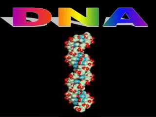
Franklin: X-ray Diffraction Patterns of DNA • British chemist, Rosalind Franklin carried out an X-ray diffraction analysis of DNA. • The diffraction patterns she obtained suggested that the DNA molecule had the shape of a helix, or corkscrew, with a diameter of about 2 nanometers and a complete helical turn every 3.4 nanometers
Watson and Crick: A Model of the Double Helix • Learning informally of Franklin’s results before they were published in 1953, James Watson and Francis Crick, two young investigators at Cambridge University. • They tried various possibilities before they finally hit on the idea that the molecule might be a simple double helix, with the bases of two strands pointed inward toward each other, forming base-pairs. • Because hydrogen bonds can form between the bases in a base-pair, the double helix is stabilized as a duplex DNA molecule composed of two antiparallel strands • The Watson–Crick model explained why Chargaff had obtained the results he had.
- + Cytosine N O O N N N N Guanine H H O N + - H - + N N N H H Base PairingGuanine And Cytosine
H H N CH3 + - Adenine Thymine O O N N N N N - N N H + Base PairingAdenine And Thymine
How does DNA replicate? • The Watson–Crick model immediately suggested that the basis for copying the genetic information is complementarity. • One chain of the DNA molecule may have any conceivable base sequence, but this sequence completely determines the sequence of its partner in the duplex. • For example, if the sequence of one chain is 5′- ATTGCAT-3′, the sequence of its partner must be 3′-TAACGTA-5′. Thus, each chain in the duplex is a complement of the other.
Three proposed models of DNA replication: • Semiconservative replication: would produce two copies that each contained one of the original strands and one new strand. • Conservative replication: would leave the two original template DNA strands together in a double helix and would produce a copy composed of two new strands containing all of the new DNA base pairs. • Dispersive replication: would produce two copies of the DNA, both containing distinct regions of DNA composed of either both original strands or both new strands.
The three hypotheses of DNA replication were evaluated in 1958 by Matthew Meselson and Franklin Stahl of the California Institute of Technology. • These two scientists grew bacteria in a medium containing the heavy isotope of nitrogen, 15N, which became incorporated into the bases of the bacterial DNA. • After several generations, the DNA of these bacteria was denser than that of bacteria grown in a medium containing the lighter isotope of nitrogen, 14N. • Meselson and Stahl then transferred the bacteria from the 15N medium to the 14N medium and collected the DNA at various intervals.
By dissolving the DNA they had collected in a heavy salt called cesium chloride and then spinning the solution at very high speeds in an ultracentrifuge. • The enormous centrifugal forces generated by the ultracentrifuge caused the cesium ions to migrate toward the bottom of the centrifuge tube, creating a gradient of cesium concentration, and thus of density. • Each DNA strand floats or sinks in the gradient until it reaches the position where its density exactly matches the density of the cesium there. • Because 15N strands are denser than 14N strands, they migrate farther down the tube to a denser region of the cesium gradient.
The DNA collected immediately after the transfer was all dense. • However, after the bacteria completed their first round of DNA replication in the 14N medium, the density of their DNA had decreased to a value intermediate between 14N-DNA and 15N-DNA. • After the second round of replication, two density classes of DNA were observed, one intermediate and one equal to that of 14N-DNA. • Thus, this experiment clearly confirmed the prediction of the Watson-Crick model that DNA replicates in a semiconservative manner.
The Replication Process • To be effective, DNA replication must be fast and accurate. • The replication of DNA begins at one or more sites on the DNA molecule where there is a specific sequence of nucleotides called a replication origin. • There the DNA replicating enzyme DNA polymerase III and other enzymes begin a complex process that catalyzes the addition of nucleotides to the growing complementary strands of DNA. • Before considering the replication process in detail, let’s take a closer look at DNA polymerase III.
DNA Polymerase III • The enzyme is a dimer, with two similar multisubunit complexes, each complex catalyzes the replication of one DNA strand. • The subunits include: • A single large catalytic α subunit that catalyzes 5′ to 3′ addition of nucleotides to a growing chain. • A smaller ε subunit that proofreads 3′ to 5′ for mistakes. • A ring-shaped β2 dimer subunit that clamps the polymerase III complex around the DNA double helix. • Polymerase III progressively threads the DNA through the enzyme complex, moving it at a rapid rate, some 1000 nucleotides per second.
The Need for a Primer • One of the features of DNA polymerase III is that it can add nucleotides only to a chain of nucleotides that is already paired with the parent strands. • Instead, another enzyme, an RNA polymerase called primase constructs an RNA primer, a sequence of about 10 RNA nucleotides complementary to the parent DNA template. • DNA polymerase III recognizes the primer and adds DNA nucleotides to it to construct the new DNA strands. • The RNA nucleotides in the primers are then replaced by DNA nucleotides.
The Two Strands of DNA Are Assembled in Different Ways • Another feature of DNA polymerase III is that it can add nucleotides only to the 3′ end of a DNA strand (the end with an —OH group attached to a 3′ carbon atom). • This means that replication always proceeds in the 5′ → 3′ direction on a growing DNA strand. • Because the two parent strands of a DNA molecule are antiparallel, the new strands are oriented in opposite directions along the parent templates at each replication fork. • Therefore, the new strands must be elongated by different mechanisms.
The leading strand, which elongates toward the replication fork, is built up simply by adding nucleotides continuously to its growing 3′ end. • In contrast, the lagging strand, which elongates away from the replication fork, is synthesized discontinuously as a series of short segments that are later connected. • These segments, called Okazaki fragments, are about 100 to 200 nucleotides long in eukaryotes and 1000 to 2000 nucleotides long in prokaryotes. • Each Okazaki fragment is synthesized by DNA polymerase III in the 5′ → 3′ direction, beginning at the replication fork and moving away from it.
When the polymerase reaches the 5′ end of the lagging strand, another enzyme, DNA ligase, attaches the fragment to the lagging strand. • The DNA is further unwound, new RNA primers are constructed, and DNA polymerase III then jumps ahead 1000 to 2000 nucleotides (toward the replication fork) to begin constructing another Okazaki fragment. • If one looks carefully at electron micrographs showing DNA replication in progress, one can sometimes see that one of the parent strands near the replication fork appears single-stranded over a distance of about 1000 nucleotides. • Because the synthesis of the leading strand is continuous, while that of the lagging strand is discontinuous, the overall replication of DNA is said to be semidiscontinuous.
The Replication Process • The replication of the DNA double helix is a complex process that has taken decades of research to understand. • It takes place in five interlocking steps: • Opening up the DNA double helix: • Stage one:Initiating replication:The binding of initiator proteins to the replication origin starts an intricate series of interactions that opens the helix. • Stage two:Unwinding the duplex:After initiation, “unwinding” enzymes called helicases bind to and move along one strand, shouldering aside the other strand as they go.
Stage three:Stabilizing the single strands.The unwound portion of the DNA double helix is stabilized by single-strand binding protein, which binds to the exposed single strands, protecting them from cleavage and preventing them from rewinding. Stage four:Relieving the torque generated by unwinding.For replication to proceed at 1000 nucleotides per second, the parental helix ahead of the replication fork must rotate 100 revolutions per second! To relieve the resulting twisting, called torque, enzymes known as topisomerases cleave a strand of the helix, allow it to swivel around the intact strand, and then reseal the broken strand.
2. Building a primer: • New DNA cannot be synthesized on the exposed templates until a primer is constructed, as DNA polymerases require 3′ primers to initiate replication. • The necessary primer is a short stretch of RNA, added by a specialized RNA polymerase called primase in a multisubunit complex informally called a primosome. • Why an RNA primer, rather than DNA? Starting chains on exposed templates introduces many errors; RNA marks this initial stretch as “temporary,” making this error-prone stretch easy to excise later.
3. Assembling complementary strands: • Next, the dimeric DNA polymerase III then binds to the replication fork. • While the leading strand complexes with one half of the polymerase dimer, the lagging strand is thought to loop around and complex with the other half of the polymerase dimer. • Moving in concert down the parental double helix, DNA polymerase III catalyzes the formation of complementary sequences on each of the two single strands at the same time.
4. Removing the primer: • The enzyme DNA polymerase I now removes the RNA primer and fills in the gap, as well as any gaps between Okazaki fragments. • 5. Joining the Okazaki fragments: • After any gaps between Okazaki fragments are filled in, the enzyme DNA ligase joins the fragments to the lagging strand.
DNA Replication Process For DNA replication some enzymes are needed: Helicase: Unwinds DNA Primase: Adds RNA primer DNA Polymerase: Adds DNA nucleotides Exonuclease: Removes RNA primer Ligase: Attaches sugar-phosphate backbone of small DNA pieces called Okazaki fragments
The particular order of the bases arranged along the sugar-phosphate backbone is calledthe DNA sequence • Genome size is usually stated as the total number of base pairs; the human genome contains roughly 3 billion bp