Download
1 / 15
150 likes | 233 Views
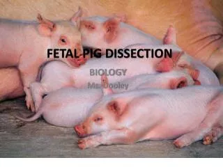
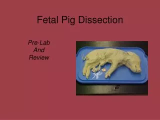
By: Derek Hu and Ben Colandrea. Fetal Pig Dissection.

E N D
By: Derek Hu and Ben Colandrea Fetal Pig Dissection
First we tied down the fetal pig with threads to ensure that it was secure and that we could enter the body without fear of instability. We used scissors and scalpel to cut through the diaphragm into the abdomen. As you can see here my partner and I have opened up the fetal pig.
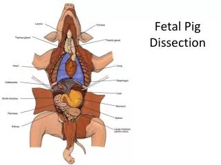
Liver Small Intestine Large Intestine Here we have a close up of the inside of the pig. Right off that bat you can see the lungs, small and large intestine, and liver.
Ribs Spleen Liver Lobes Small Intestines Here we have pulled up the liver to expose the lobes of the liver, and you can clearly see again the large and small intestines and the spleen. Also if you look closely to the side you can see where we cut into the ribs.
Stomach Umbilical Cord Kidney Here we pulled aside the intestines to reveal the kidneys, umbilical cord and stomach.
Here we have removed the intestines. If you were to unravel the whole thing it would be over 20 feet long! We used scissors to cut away the bottom of the intestines to remove it from the rest of the pig.
Here is the liver and the lungs of the pig that we have removed.
This is the heart of the pig. It resembles that of a human heart you can see the shape is similar and you can point out where the atrium and ventricles are.
Kidneys Spine Here we have removed most of the digestive system leaving visible structures like the spine and kidneys intact.
Finally we moved to removing the brain. After carefully making an incision in the skull we removed the top skin to reveal the cranium which covers the brain giving protection.
The cranium is actually quite tough to cut through and took a bit of force to get through but we removed most of it to reveal the cerebral cortex of the brain.
Careful cutting allows for the brain to simply slip out of the brain cavity and here is the pig’s brain shortly after removing it. Notice the distinct brain like shape and the visible similarities with lobes.
Brain Stem connects here with spine. Finally here is a picture of the pig’s head with the brain removed, the brain fits snuggly into the cavity and you can see where the brain stem is attached to which connects the brain with the spine and the whole nervous system allowing the pig to function.
Mobile Devices To aid us in our project we used a first generation iPad to help us to understand the anatomy of the pig. We also used my camera, a digital camera to take pictures of the inside of the pig.
We used the iPad to go onto the website http://www.whitman.edu/content/virtualpig. This provided a detailed walkthrough through the dissection and helped my partner and me to identify the different organs. In future lessons the iPad is an invaluable tool because it allows to quickly check one’s progress during the dissection. My camera was used to take high resolution pictures of parts of the pig and then I incorporate these pictures into my PowerPoint. Without pictures it would be difficult to simply describe how parts of the pig looked with words.