Download
1 / 32
1.27k likes | 3.14k Views
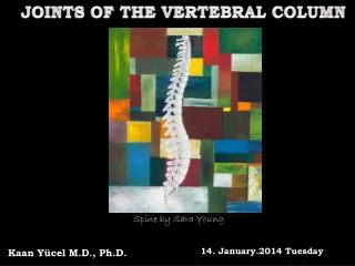
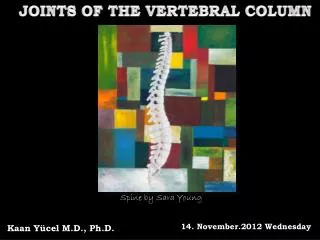
Anatomy of the vertebral column. Objectives. Describe the general structure of the vertebra Describe the structure of the atypical cervical vertebrae Describe the structure of typical cervical vertebrae Describe the structure of the thoracic vertebra

E N D
Objectives • Describe the general structure of the vertebra • Describe the structure of the atypical cervical vertebrae • Describe the structure of typical cervical vertebrae • Describe the structure of the thoracic vertebra • Describe the structure of the lumbar vertebra • Describe the structure of the sacrum • List the differences between vertebrae in different regions of the vertebral column
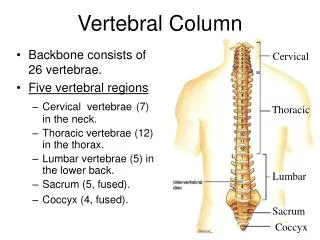
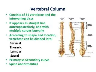
Vertebral Column • Consists of 31 vertebrae and the intervening discs • It appears as straight line anteroposteriorly, and with multiple curves laterally • According to shape and location, vertebrae can be divided into: Cervical Thoracic Lumbar Sacral • Primary vs Secondary curve • Spine abnormalities
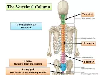
Vertebral Column • 7 C, 12 T, 5 L, 5 S (Fused as Sacrum), 4 coccygeal • Primary Curves • Secondary Curves
Typical Vertebrae • Body • Superior and inferior surfaces of body (plateaus) • Thickened around the rim, location of epiphyseal plates • Cartilaginous end-plates • Vertebral Arch • Pedicles, Laminae • Transverse Processes • Spinous Process • Facets – superior articular and inferior articular • Vertebral Foramen • Vertebral notch • Intervertebral Foramen
Ligaments of the Spine • Anterior longitudinal • Posterior longitudinal • Supraspinous • Interspinous • Ligamentumflavum
Intervertebal Discs • Intervertebral Discs • Fibrocartilaginous joints • Increase in size from C to L (3mm to 9 mm) • Make up 20-30% of length of column
Discs • Outer rim of fibrocartilage called the anulusfibrosus(attaches to cartilaginous end plate) • Connects vertebral bodies in a fibrocartilaginous joint (no capsule, little motion) • Anulus encloses a central mass called the nucleus pulposus • About 80-90% water, less with increased age • Contains a mucopolysaccharide matrix • Changes shape, releases and absorbs water. • Neither blood vessels or nerves penetrate nucleus
Discs • Structure deforms when pressure is put on vertebral column as in weight bearing • Acts as a shock absorber • Annulus totally encloses the nucleus and keeps it under constant pressure • With increased age, the H2O content decreases and the nucleus becomes more fibrocartilaginous, therefore less easily deformable and more easily damaged
Discs • Nucleus, when under extreme pressure, can herniate or extrude from the disc in a posterior or posterior-lateral direction • Usually occurs in cervical or lumbar region • Nucleus can put pressure on spinal nerve causing referred symptoms (motor and sensory) • Can cause pressure on cord itself if true posterior
Cervical Vertebrae • 1st cervical vertebra(Atlas) No body No spine Anterior arch Posterior arch • 2nd cervical vertebra (Axis) looks like typical vertebra It has odontoid process (Dens)
Typical Cervical Vertebrae • From C4 to C7: Characterized by the general structure of vertebra but each has a bifid spine • 7th cervical vertebra: Characterized by general structure of vertebra but it has NO bifid spine
Thoracic Vertebra • Heart-shaped body • Long, thin, vertical spine • Round vertebral foramen • Body/foramen ratio almost 2 • Superior articulating facets facing posteriorly • Body has impression for rib articulation( COSTAL facet)
Lumbar Vertebra • Kidney-shaped body • Short, thick, horizontal spine • Triangular vertebral foramen • Body/foramen ratio almost 3-4 • No impression for rib articulation
The Sacrum • Identify the following: Anterior sacral foramina Transverse lines Promontory Ala Posterior sacral foramina Median sacral crest Medial (Intermediate) sacral crest Lateral sacral crest Sacral hiatus Cornu Sacral canal
Atlanto-Occipital Joint • Two concave superior facets of atlas articulate with two convex surfaces of occipital condyles of the skull • Supported by major ligaments: 1- Ant. Atlanto-Occipital, 2- Tectorial Membrane, 3- Post. A-O • Small saddle joint • Very limited motion – • nodding type motions in all directions.
Atlanto-Axial Joint • Atlas and Axis • Pivot • Two convex superior facets of axis with two concave inferior facets of the atlas • Atlas also posses a facet on the internal surface of the anterior arch which articulates with the dens of the axis • Major ligaments from spine support – • Ant. Atlanto-Occipital, • Tectorial Membrane, • Post. A-O