Download
1 / 36
500 likes | 2.52k Views
Methods in Microbial Ecology Lecture 16 Feb 27, 2006 The study of how microorganisms interact with each other and with their environment. Biodiversity – isolation, identification and quantification of microorganisms in their native habitats

E N D
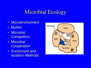
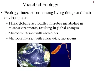
The study of how microorganisms interact with each other and with their environment. • Biodiversity – isolation, identification and quantification of microorganisms in their native habitats • Microbial activity – what are the organisms actually doing in their native habitats Microbial ecology
In nature, microorganisms exist in mixed cultures called microbial communities • To isolate and study a particular organism, researchers must isolate microorganisms from nature and establish them in pure cultures • Enrichment culture is a selective medium and incubation procedure that selects for desired organisms and against undesired ones. Success requires a proper inoculum, culture medium and incubation conditions Culture-dependent analysis
Enrichment and isolation of Azotobacter • Azotobacter is a fast-growing nitrogen-fixing bacterium • It is isolated in culture conditions including oxygen, and no fixed nitrogen supply
A miniature anoxic ecosystem that is used as a long-term inoculant source for enrichment cultures • Particularly good for enrichment of purple and green phototrophic bacteria, sulfate reducing bacteria • Preparation – 1) fill a large glass cylinder half full with organic-rich mud supplemented with calcium carbonate and calcium sulfate. 2) Top up with lake, pond or sea water. 3) Cap loosely and grow in muted sunlight Winogradsky column
Test the hypothesis that a particular organism exists in the column by supplementing with a compound that it can degrade
A mixture of organisms generally develops: • Algae and cyanobacteria appear in the upper part of water column and produce oxygen • Anoxic decomposition in the mud creates a gradient of hydrogen sulfide which favors growth of green and purple sulfur bacteria • Purple nonsulfur bacteria may grow at the interface between oxic and anoxic zones
Dominant organism in an enrichment culture does not necessarily represent the dominant organism in a natural habitat. • Laboratory cultures often misrepresent natural communities. Lab cultures favour fastest-growing species • Dilution of the inoculum prior to enrichment often yields a different culture. Why? Enrichment bias
Dilution of a mixed culture in tubes containing molten agar. Individual colonies become separated and embedded in the agar • Useful for purifying anaerobes such as phototrophic sulfur bactera and sulfate-reducing bacteria from Winogradsky columns. • Pure cultures are generally obtained after several serial dilutions Pure culture isolation – agar shakes
Pure culture isolation – Most Probable Number technique • Serial dilution of inoculum in liquid medium until the final tube shows no growth. • Used to test numbers of microorganisms in foods, wastewater and other samples where cell numbers are measured routinely • Medium can be highly selective for individual species or complex enough to get an idea of total cell numbers
MPN in this example is 105 – 106 recoverable cells per gram of sample
High-tech pure cultures-Laser tweezers • Apparatus consists of an inverted light microscope, powerful infrared laser and micromanipulation device • Laser creates a force pushing down on small objects (such as a single cell) that can be moved away from contaminants • Useful for isolating slow-growing or rare species
Cells in capillary tubes can be optically trapped, isolated. Tube is broken and the cell is flushed out into sterile medium to initiate a pure culture
Molecular analysis – viability and quantification staining • DAPI – 4’,6-diamido-2-phenylindole is a fluorescent stain useful for quantitating cells in opaque habitats • DAPI binds to DNA and fluoresces bright blue, making cells easy to count • Aquatic samples can be counted after the sample is passed through a filter • DAPI staining does not distinguish living from dead cells or between different microbial groups
Methods of fluorescent staining that distinguish between living and dead cells. Example: LIVE/DEAD Bac Light • Based on the integrity of the cytoplasmic membrane • Green fluorescent dye penetrates all cells. Red dye containing propidium iodide penetrates only those cells with severely damaged membrane. Red cells = dead cells • Procedure does not work in natural habitats because of nonspecific fluorescence of background materials Viability staining
Exploits the specificity of antibodies to recognize a particular cell-surface protein of an organism • Used for identifying an organism in a complex community including many species, such as soil or a clinical sample • Preparation of antibodies against a particular organism is expensive and time-consuming • Many clinically relevant antibodies are available commercially Fluorescent antibodies-immunofluorescence
Detection of Sulfobolus acidocaldarius on the surface of soil particles by immunofluorescence
Limitations of cell staining/microscopy • Very small cells may be overlooked • Morphologically similar but genetically distinct species may be confused • Genetic stains solve this problem by detecting specific gene sequences that can be quite specific, or quite promiscuous
Fluoresent in situ hybridization uses fluorescently-labelled single-stranded DNA or RNA probes to bind directly to its complementary sequence in a nucleic acid. • Phylogenetic staining using FISH uses probes that bind directly to ‘signature’ sequences within 16S (prokaryotes) or 18S (eukaryotes) rRNA. Degree of specificity of the probes can be altered to detect individual species or entire domains. Can therefore be used to track related organisms Genetic Stains - FISH
FISH technology used to detect the presence of specific genes in a sample. Example – is a nitrogen-fixing organism present? Look for nitrogenase • Must have a fluorescent probe specific for the class of genes of interest – can distinguish photosynthetic organisms, nitrogen fixers, hydrogen bacteria etc. • Used to estimate the numbers of different types of cells in a natural sample Chromosome painting
Used to determine whether a given gene is being expressed in a sample at a particular time • Involves the use of a probe that binds to the mRNA of the gene of interest. Therefore only the expressed gene is detected • Allows researchers to study factors affecting gene expression in natural populations and habitats In situ reverse transcription
PCR – linking genes to specific organisms • Biodiversity of a habitat can be monitored without culturing or observing cells. Isolate characteristic gene sequences instead! • Isolate total DNA from habitat and clean up contaminants. Use PCR to amplify gene sequences of interest (often 16S RNA) • Sequence individual PCR products or separate them by denaturing gradient gel electrophoresis
The most abundant members of a microbial community may never have been seen in laboratory culture
Measuring microbial activity in nature-radioisotopes • Direct chemical measurements are often sufficient to detect microbial activities – transformation of lactate and sulfate to hydrogen sulfide by sulfate-reducing bacteria • Radioisotopes are used to determine the fate of portions of molecules, turnover rate, or for extreme sensitivity
Controls such as the ‘killed cell control’ guarantee that transformation of a radiolabeled compound is due to a microbial rather than a strictly chemical process
Tiny glass electrodes (2-100 m diameter) to measure pH, oxygen, carbon dioxide, hydrogen, hydrogen sulfide. Often used to measure chemical reactions in microbial mats: • Layered microbial communities with cyanobacteria in the uppermost layer, anoxygenic phototrophs in middle layers and chemoorganotrophs (especially sulfur reducing) as light becomes limiting at the bottom • Microbial mats are found in hot springs and intertidal zones Microelectrodes
Isotopes that are not radioactive. Carbon and sulfur are the most commonly used in microbiology • 12C is 95%, 13C is 5% • 32S is common, 34S is rare • Biochemical reactions tend to favour the lighter isotope. Therefore cells become enriched in 12C and depleted in 13C relative to a standard. Stable isotopes
Isotopic fractionation – discrimination against the heavier isotope
Use of isotopic fractionation in microbial ecology • Isotopic composition of a sample indicates its past biological activity • Life can be inferred from organic carbon in rocks 3.5 billion years old • Sulfides in lunar rocks provide evidence against past life on the moon