Download
1 / 14
140 likes | 150 Views
Techniques to Study the Brain. 3B: The Brain. How do neuroscientists study the brain’s connections to behavior and mind?. In the olden days…. In the past how did we study brain anatomy?

E N D
3B: The Brain • How do neuroscientists study the brain’s connections to behavior and mind?
In the olden days… • In the past how did we study brain anatomy? • It was really difficult to study brain anatomy and function unless somebody ________ or an __________ occurred. • Luckily, with modern technology,: • We don’t necessarily have to wait for tragedy to get our answers!
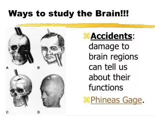
Studying the Brain: Research Methods • Clinical observations • Damage studies/lesioning • Electroencephalography (EEG) • Electrical stimulation (ESB) • Brain imaging – • computerized tomography (CT “Cat” scan) • positron emission tomography (PET scan) • magnetic resonance imaging (MRI)
Clinical Observation • Clinical observations have shed light on a number of brain disorders. • Alterations in brain morphology due to neurological and psychiatric diseases are now being catalogued.
Lesions • A brain lesion involves experimentally __________ and ________ sections of brain in animals, then observing their behaviors after such destruction Hubel (1990)
Electroencephalogram (EEG) • An amplified recording of the electrical waves (neural activity) sweeping across the brain’s surface, measured by electrodes placed on the scalp.
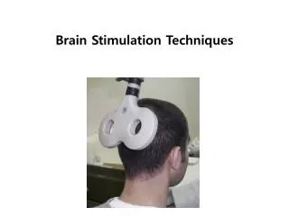
Electrical Stimulation (ESB) • Researchers electrically stimulate parts of the brain during brain surgery and note the effects • Applying currents that can shut down or activate certain parts of the brain • Can you think of any diseases or conditions this could possibly help fix?
Deep Brain Stimulation Surgery • Deep Brain Stimulation Surgery: • http://www.youtube.com/watch?v=w8zPQ8YV43I • http://www.youtube.com/watch?v=q7_7bwHmG4M • http://www.youtube.com/watch?v=j3NjNKm0pio • http://www.youtube.com/watch?v=Lq5rIILcVgA
Computerized Tomography (CT or “Cat” scan) • CT scanning combines special x-ray equipment with sophisticated computers to produce multiple images or pictures of the inside of the body. • These cross-sectional images of the area being studied can then be examined on a computer monitor or printed. • Provides greater clarity and more details than regular x-ray exams.
Positron Emission Topography (PET Scan) • PET (positron emission tomography) Scan is a visual display of brain activity that detects a radioactive form of glucose (sugar) while the brain performs a given task. • Active areas have increased • blood flow • Sensors detect • radioactivity • Different tasks show • distinct activity patterns
Magnetic Resonance Imaging (MRI) • An MRI is basically a picture of the brain from many angles and it provides clear 3D images • It uses magnetic fields and radio wavesto produce computer-generated images that distinguish among different types of brain tissue. • The images to the right show ventricular enlargement in a schizophrenic patient.
Functional MRI (fMRI) • Don’t confuse an MRI with a functional MRI • A “functional” MRI enables doctors to see actual movement and activity in the brain. MRI just provides detailed pictures of the brain. • The image to the right shows brain regions that are active when a participant lies.