Download
1 / 28
420 likes | 660 Views
CLINICAL ANATOMY OF ORAL CAVITY. By Dr.Sanaa Alsharawy. Objectives and Clinical Significance of the Oral Cavity :. The mouth is one of the important areas that the medical professional is called on to examine,so you should :

E N D
CLINICAL ANATOMY OF ORAL CAVITY By Dr.SanaaAlsharawy
Objectives and Clinical Significance of the Oral Cavity : The mouth is one of the important areas that the medical professional is called on to examine,so you should : Knew the nerve supply, blood and lymph drainage of the mouth cavity contents. Evaluate the movement of soft palate. Examine the oral part of pharynx for tonsilitis. Knew the close relation of the submandibular duct and calculus formation. Remember the close relation of the lingual nerve to the lower 3rd molar tooth and to the submandibular duct. Evaluate the mucous membrane and movement of the Tongue. Identify the clinical estimation of hypoglossal nerve.
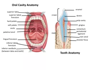
ORAL CAVITY • The mouth : • It is divided into the 1- Vestibule: • Which lies between teeth & gums internally and cheeks &lips externally. • The parotid duct opens opposite the upper second molar. • 2- Mouth cavity proper: • Which lies within the alveolar arches, teeth and gums. Vestibule Mouth Proper
MOUTH • Mouth proper:has a roof,which is formed by thehard & soft palate. • Thefloor is formed by the anterior 2/3 of the tongue
PALATE • The palate forms the roof of the mouth. • It is divided into two parts: • The hard (Bony) palatein front and • The soft palate behind.
HARD PALATE HARD PALATE • The hard palate is formed by (4 bones), palatine processes of the maxillae and horizontal plates of palatine bones. • It forms the floor of the nasal cavities and the roof of the mouth cavity.
SOFT PALATE • It is a mobile fold of mucous membrane filled with striated muscles. • It is attached to the posterior border of the hard palate. • Its free posterior end is a conical projection called the uvula.
Sensory innervation of Soft Palate • The greaterandlesser palatine nervesfrom the maxillary nerve. • The nasopalatine nerve,also a branch of themaxillarynerve. • The glossopharyngeal nerve also supplies the soft palate.
BLOOD SUPPLY OF THE PALATE • Greater & lesser palatine branches of the maxillary artery. • Ascending palatinebranch of the facial artery. • Ascending pharyngeal branchof the external carotid artery.
MUSCLES OF THE SOFT PALATE 5 pairs of muscles 1-Tensor veli palatini, 2- Levator veli palatini, 3- Palatoglossus, 4- Palatopharyngeus, 5- Musculus uvulae.
MOTOR INNERVATION OF SOFT PALATE • All muscles are supplied by pharyngeal plexus EXCEPT the tensor vili palatini is supplied bynerve to medial pterygoid muscle from MANDIBULAR NERVE.
It is a network of nerve fibers innervating most of the palate, pharynx and larynx. • It lies on the outer wall of pharynx, mostly on the midlle constrictor. • It is formed of pharyngeal branches of glossopharyngeal N.,vagus N. including fibres of Cranial root of accessoryand superior cervical sympathetic ganglion. Pharyngeal plexus
Clinical Estimation of MOVEMENTS OF SOFT PALATE • Clinically , Motor innervation of soft palate Can be tested by saying ‘ah’, Normallysoft palate rises and uvula moves backward in the middle. • Pharyngeal isthmus (the communication between nasal and oral parts of the pharynx) is closed by raising the soft palate via contraction of levatorpalatini. Closure occurs during the production of explosive acts in speech & in swallwing.
Clinical Significance of the Oral part of Pharynx • The palatine tonsils are two masses of lymphoid tissue located in lateral walls of the oral part of pharynx in the tonsillar sinuses. • The palatine tonsils are the common site of infection, producing the characteristic tonsilitis. • The deep cervical lymph node, which situated below and behind the angle of mandibleis usually enlarged and tender. • Recurrent attacks of tonsilitis are treated by tonsillectomy. • Clinically, the external palatine vein, which lies lateral to the tonsil, may be the source of postoperative bleeding.
TONGUE • The tongue is a mass of striated muscles covered with mucous membrane. • Its anterior 2/3 lies in the mouth, and its posterior 1/3 lies in the pharynx. • It has several important functions: • Normal articulation of the jaw. • Manipulation of food and swallowing. • Production of normal speech.
Mucous Membrane of tongue Tongue • The upper surface (Dorsum)of the tongue can be divided into anterior 2/3 or oral part and/ posterior 1/3 or pharyngeal part by a V-shaped sulcus.Thesulcus terminalis. • The apex of the sulcusis marked by a small pit, the foramen cecum. • It is Embryologic remnant of the upper end of the thyroglossal duct.
Mucous Membrane of tongue • Three types of papillae are present on the upper surface of the anterior two thirds of the tongue: the filiformpapillae, thefungiformpapillae, and the vallate papillae. • The mucous membrane covering the posterior third of the tongue is devoid of papillae but has a nodular irregular surface caused by the presence of underlying lymph nodules, the lingual tonsil.
Changes in the mucosa lining the tongue indicate thesystemic diseases, such as diabetes or vitamin deficiency, or the local effects of chronic tobacco or alcohol use.
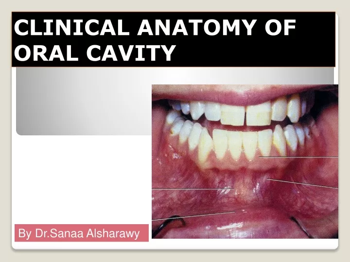
Floor of the Mouth • Mucous membrane on the under surface of the tongue is smooth. • In the midline, the undersurface of the tongue is connected to the floor of the mouth by a fold of mucous membrane, the frenulum of the tongue. • The submandibular duct opens into the floor of mouth on each side of frenulum of tongue. • The sublingual ducts also open by many openings (8-20) into the floor of mouth on the summit of sublingual fold on the lateral side of submandibular duct opening.
Calculus formation : • It is a tense swelling below the body of the mandible,which is greatestduring a meal and is reduced in size or absentbetween meals(diagnostic of the case). • Clinically: • by examination of floor of mouth, reveals absence of ejection of salivafrom the orifice of duct.+ stone can be palpated in theduct, which lies below m.m. of the floor of mouth. • During the operation, we should remember that the duct is crossed by the lingual nerve. Clinical Anatomy of Submandibular Duct
MUSCLES OF THE TONGUE • The muscles of the tongue are divided into two types: • Intrinsicand extrinsic. • The intrinsic muscles are restricted to the tongue and are not attached to bone. • They consist of longitudinal, transverse, and vertical fibers. • Nerve supply:Hypoglossal nerve. • Action:Alter the shape of the tongue while it lies in the mouth cavity.
Extrinsic Muscles of the Tongue • The extrinsic muscles are 4 pairs attached to bones and the soft palate. • They are: • Palatoglossus. • Styloglossus, and • Genioglossus, • Hyoglossus. All muscles of the tongue are supplied by the hypoglossal nerve EXCEPTpalatoglossus which is supplied by the pharyngeal plexus
SENSORY INNERVATION • General sensations from the anterior 2/3of the tongue are carried by the lingual nerve. • Taste fibersfrom the anterior 2/3 excluding the vallate papillae, are carried by thechorda tympaniof the facial nerve. • General & taste sensationsfrom the posterior 1/3 ,including the vallate papillae, are carried by the glossopharyngeal nerve. • General & taste sensations from root of the tongue and epiglottis are carried by the vagus nerve.
Injury to Lingual nerve • The dangerous area during tooth extraction; • Here, Lingual nerve is closely related to the lowerlast molar tooth and is liable to be damaged in cases of clumsy extraction on an impacted 3rd molar.
Blood Supply • 1-The main artery is the lingual artery • 2- Tonsillar branch of the facial artery, • 3- Ascending pharyngeal artery. • The veins drain into the internal jugular vein. External Carotid Artery
LYMPHDRAINAGE • The tip of the tongue drains into the submentallymph nodes. • The remainder of the anterior 2/3 of the tongue drains into the submandibular& deep cervicallymph nodes. • Lymph from the posterior 1/3 of the tongue drains into the deep cervicallymph nodes. • The lymphatic drainage is important in the early spread of carcinoma of the tongue.
Ask the patient to protrude his tongue : • Normally, Rt.&Lt. genioglossus muscles contract together protruding the tip of tongue anteriorly in the middle line. • Lesion of hypoglossal N. on Rt. side leads to atrophy & wrinklingof the tongue on the same side of lesion. • Asking patient to protrude the tongue, the tip deviates to side of the lesion. Clinical Estimation of the Hypoglossal Nerve