Download
1 / 18
180 likes | 219 Views
Epithelial Tissue. Objectives: By the end of this lecture, you should be able to: Describe general characteristics of epithelial tissue. Discuss microscopic structure and distribution of different types of epithelial membranes.

E N D
Epithelial Tissue Objectives: By the end of this lecture, you should be able to: • Describe general characteristics of epithelial tissue. • Discuss microscopic structure and distribution of different types of epithelial membranes. • Classify glandular epithelium according to different parameters. • Enumerate the functions of epithelial tissue. • Understand the following clinical applications: • Immotile cilia syndrome (Kartagener’s syndrome). • Metaplasia.
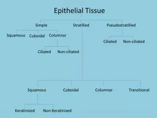
EPITHELIAL TISSUE General characteristics: • Cells are tightly joined withlittle intercellular space. • Rest on a basement membrane. • Avascular. • High power of regeneration. Classification: • Epithelial membranes: • Simple epithelium: one layer. • Stratified epithelium: more than one layer. • Glands (Glandular Epithelium).
I. Simple Epithelium 1- Simple Squamous Epithelium: One layer of flat cells with flat nuclei. Provides smooth thin surface. Examples of sites: • Endothelium (lining the CVS “cardiovascular system”). • Alveoli“air sacs”of lung.
I. Simple Epithelium 2- Simple Cuboidal Epithelium: One layer of cuboidal cells with central rounded nuclei. Example of sites: • Thyroid follicles.
I. Simple Epithelium 3- Simple Columnar Epithelium: • One layer of columnar cells with basal oval nuclei. • Types: • Non-ciliated:Example of sites: Lining of stomach, gall bladder, and intestines (with goblet cells). • Ciliated: with cilia on free surface.Example of sites: Fallopian tubes.
I. Simple Epithelium 4- Pseudo-Stratified Columnar: • One layer of columnar cells. • Some cells are tall. • Others are short and don’t reach the surface. • All cells rest on the basement membrane. • Nuclei appear at different levels. • Types: • Non-ciliated:Example of sites: vas deferens. • Ciliated with Goblet Cells “respiratory epithelium”:Example of sites: trachea & bronchi.
II. Stratified Epithelium 1- Stratified Squamous Epithelium: • Multiple layers of cells. • Basal cells are columnar with basal oval nuclei. • Intermediate cells are polygonal with central rounded nuclei. • Surface cells are flat with flattened nuclei. • Types: • Keratinized: with a layer of keratin on the surface.Example of sites: epidermis of skin. • Non-keratinized: without a layer of keratin on the surface.Example of sites: esophagus.
II. Stratified Epithelium 2- Transitional Epithelium: • Multiple layers of cells. • Basal cells are columnar. • Intermediate cells are polygonal. • Surface cells large cuboidal with convex free surface and may be binucleated. • Example of sites: Urinary bladder.
II. Stratified Epithelium 3- Stratified Columnar Epithelium: • Multiple layers of cells. • Basal cells are columnar. • Intermediate cells are polygonal. • Surface cells are columnar. • Example of sites: large ducts of glands.
GLANDS (Glandular Epithelium) Classification: 1- According to presence orabsence of ducts: a. Exocrine:e.g. salivary glands. b. Endocrine:e.g. thyroid gland. c. Mixed: e.g. pancreas. 2- According to number of cells: a. Unicellular: e.g. goblet cells. b. Multicellular: e.g. salivary glands.
GLANDS (Glandular Epithelium) Classification: 3- According to mode of secretion: a. Merocrine: No part of the cell is lost with the secretion, e.g. salivary glands. b. Apocrine: The top of the cell is lost with the secretion, e.g. mammary gland. c. Holocrine: The whole cell detaches with the secretion, e.g. sebaceous glands. Mero-crine Apo-crine Holo-crine
GLANDS (Glandular Epithelium) Classification: 4- According to shape of secretory part: 1. Tubular: e.g. intestinal gland. 2. Alveolar (acinar): e.g. mammary gland. 3. Tubulo-alveolar: e.g. pancreas.
GLANDS (Glandular Epithelium) Classification: 5- According to nature of secretion: a. Serous:e.g. parotid gland. b. Mucous: e.g. goblet cells. c. Muco-serous:e.g. sublingual gland. d. Watery: e.g. sweat gland.
FUNCTIONS OF EPITHELIUM 1- Protection as in epidermis of skin. 2- Secretion as in glands. 3- Absorption as in small intestine. 4- Excretion as in kidney. 5- Reproduction as in gonads. 6- Smooth lining as in blood vessels.
Clinical Applications • Immotile cilia syndrome (Kartegener’s syndrome): • Disorder that causes infertility in male and chronic respiratory tract infection in both sexes. • It is caused by immobility of cilia and flagella induced by deficiency of dynein. • Dynein protein is responsible for movements of cilia and flagella.
Clinical Applications • Metaplasia: • It is the transformation of one type of tissue to another in response to injury. This condition is usually reversible if the injury is removed. • Example: pseudostratified ciliated columnar epithelium of the respiratory passages, e.g. trachea, of heavy smokers may undergo squamous metaplasia, transforming into stratified squamous epithelium.