Download
1 / 69
830 likes | 2.41k Views
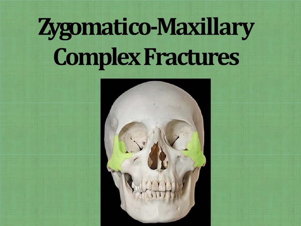
Z y g om a ti c o -M a xilla r y C omplex Fracture s. Introduction. 2 nd most common facial fractures after nasal fractures . High incidence is because of prominent position with i n facial skeleton. Male : female= 4:1 Peak incidence: 2-3 decade Left ZMC # > Right

E N D
Introduction 2nd most common facial fractures after nasal fractures. Highincidenceis becauseofprominentpositionwithinfacialskeleton. Male : female=4:1 Peak incidence: 2-3decade Left ZMC # > Right Bilateralfractures are rare= 4% Affects facial contour, ocular andmandibularfunctioning.
Fracture of zygoma is not usually present alone,itsfindmostlyintheconjunctionwith adjacent structurei.e. • antrum, • orbital walls including the infraorbitalcanal, • rim • orbitalfloor • This structure makes up the zyogmaticomaxillary complex
AppliedAnatomy It is roughly equivalent of a four sided pyramid sidesare represented by fourprocesses Temporal Orbital Maxillary Frontal
Articulations • Four articulatingsutures: • Zygomatico-frontal • Zygomatico-temporal • Zygomatico-maxillary • Zygomatico-sphenoid • In a series of 134 zygoma fractures at the District of Columbia General Hospital, the zygomaticotemporal suture line on the archwas fractured most frequently, followed byfractures of the suture line on the infraorbital rim and then by the zygomaticofrontal and zygomaticomaxillary suturelines
Fracture patterns Fracture medial to a zygomatico-maxillary suture and along a zygomatic-cosphenoid suture withinorbit. Fractures through a fronto-zygomatic suture and posterior to a zygomaticotemporalsuture. Fracturesextending from theinferior orbitalfissuresuperiorly through the zygomatico-sphenoid suture and inferiorly throughthe zygomaticbuttressof the maxilla. 4. Triple fracture through the zygomaticarch.
Axis ofrotation • Vertical axis : Line passing through frontozygomatic suture and first molartooth • Medial or lateralmovement • Horizontal Axis : infraorbital foramenand horizontalarch • Upward or downward movement ofbroken segment
Rowe’s and Killey classification(1968) • Type I : no significantdisplacement • Type II : fracture of zygomaticarch • Type III : rotation arround horizontal axis – inwardor outwarddisplacement • Type IV: rotation around longitudinal axis – medialor lateral • Type V : displacement of the complex block– medial/inferior/lateral • Type VI: displacement of orbitoantralpartition • Type VII: displacement of orbital rimsegment • Type VIII: complex comminutedfracture.
ROWE AND WILLIAMS(1985) • Fractures stable afterelevation • Arch only (mediallydisplaced) • Rotation around the verticalaxis • medially • laterally
Fractures unstable afterelevation • Arch only (inferiorlydisplaced) • Rotation around horizontalaxis • medially • laterally • c) Dislocations enbloc • inferiorly • medially • postero-laterally • d) Communitedfractures
INSPECTION • Symmetry • Pupillarylevel • Presence of orbitaloedema • Sub-conjunctivalecchymosis • Ant&latprojectionofzygomaticbodies • Intra-oral • PALPATION • Systematic&thorough • Compare one side withanother
Clinical features: Skeletaldeformities • Asymmetry ofthe midface • Depression/flattening of the malar prominence • Flattening, hollowing (bony indentation)or broadening over the zygomaticarch • Palpable step offsor gap deformities of orbital margins (infraorbital/lateral)
Flattening of the MalarProminence. A characteristic sign and striking feature -flatteningofthenormalprominenceinthemalar area. Seen in especially those in which distraction of the frontozygomatic suture and medial rotationand/or comminution haveoccurred. Reported in 70% to86% ofcases. Observed in birds and wormsview.
Deformity at the Zygomatic Buttress of the Maxilla. Intraoral palpation frequently reveals irregularities of the normallysmooth contour, especially in the area of the zygomatic buttress of the maxilla. Deformity of the Orbital Margin: Fractures running through the orbitalrimoftenresultinagap,orstepdeformity,ifdisplacement hasoccurred.
Crepitation from AirEmphysema: Can be palpated by alternatively rolling two fingers gently over thetissue, which produces a characteristic cracklingsensation. Thesofttissueoftheperiorbitalarea,especiallytheeyelids,is pronetoinflation withair because of its loose areolar nature. Disappears spontaneously in 2 to 4 days withouttreatment.
Periorbital Ecchymosis and Edema: Edema and bleeding into theloose connective tissue of the eyelids and periorbital areas is the most commonsign followingfractureoftheorbitalrim
Subconjunctival Ecchymosis: Subconjunctival haemorrhage reported in 50% to 70% ofcases. It will not have posteriorlimit It will be bright red because of the ability of oxygen to diffusethrough the conjunctiva to the collectionof blood.
Unequal PupillaryLevels. Withthedisruptionoftheorbitalfloorandlateralaspectoftheorbitcauses lossofosseoussupportfortheorbitalcontentsanddisplacementofTenon’s capsuleandthesuspensoryligamentsoftheglobepermitdepressionofthe globe
Diplopia(blurredvision) Monoculardiplopia,orblurringofvisionthroughoneeyewiththeotherclosed usually indicates a detached lens, hyphema, or other traumatic injury to the globe. Binoculardiplopia:Blurringofvisionoccursonlywhenthepatientlooksthrough both eyes simultaneously. Reported in 10% to 40% of zygomaticinjuries. Causes: Muscle entrapment: doesnt resolve by itself. Checked by forced ductiontest. Edema or hemorage: Resolve in fewdays.
Diplopia:cause • Haeomotomaor edema arround extraoccular muscle • Neuromuscular injury • Disruption of attatchment of inferior rectus or inferior obliquemuscle
Diplopia :test • Finger Gaze: finger moved in front of eye inall nine direction of gaze at a distance 30 cm. Reporting of double vision –positive. • Forcedductiontest: differentiate between permanent and temporary diplopia –carried out under topicalLA. • Tendons of inferior rectus muscle is heldwith tissue holdingforceps and eyeball is rotated superiorly with othermovement • failure to rotate indicates paralysis orentrapment of muscle infracture
Enophthalmos. Increase in orbital volumeduetolateral and inferiordisplacement of thezygomaand/ordisruption of the orbital walls causes herniation of orbital resulting intoenophthalmos. Reported in 5 %cases.
Exophthalmos • Rare • Decrease in orbital volume (inward fracture of orbital walls) • Retrobulbarhemorrhage
Abnormal NerveSensibility: Infraorbital nerve paresthesia is more common in fractures thatare displaced than nondisplaced. A related symptom is altered sensitivity of the maxillaryteeth andgingiva. Reported in 50% to 90% ofZMC injuries.
Sensory alterations • Sensory deficit (hypoesthesia, anesthesia) in the distribution of: • 1. Infraorbitalnerve: lower eyelid, upperlip, ala and lateral sidewall of thenose Zygomatiofacial nerve: malar eminence,cheek Zygomaticotemporal nerve: lower lateral orbital rim anterior temporal/lateral/frontal region 3 2 1
Oralfindings • Ecchymosis of the gingivobuccal maxillarysulcus • Palpable step deformity at the buttress & antral walls • Crepitus • Subjective occlusal disorder due to altered sensation of the maxillary premolars/molars and gingiva, no objectivemalocclusion • Trismus
Trismus: • Limitation of mouth opening in approximately one third of cases. 45% inarch cases. • Impingementofthe coronoid process due to thedisplaced zygomaticfragments requires large displacements. • Therefore more accepted theory saystrismusisdue to muscle spasm rather than direct contact withcoronoid.
Epistaxis: Whenever the sinus mucosa is disrupted, haemorrhage into thesinus ispossible. Because the maxillary sinus drains into the nose via the middle meatus, unilateral hemorrhage from the noseis possible. It is reported in 30% to 50% ofZMC injuries.
WATERS VIEW :Waters’ view is the most useful view to evaluate for zygomaticinjuries because it defines the injury involving the arch, lateral wall of the maxillary sinus, inferiororbitalrim,andflooroftheorbit
Jug handleview It is essential for demonstrating the status of the zygomatic arch,posterior displacement, and lateral or medial rotation on the verticalaxis. SMV with 30% lessexposure
SurgicalApproach • Extra-oralapproach • Bicoronal orhemicoronal • Gillies :Temporal • Superolateral • Supraorbital approach : lateraleyebrow • Uppereyelid • Lowereyelid • Infraorbital • Subtarsal • Subcilliary/infracilliary : lowerblepheroplasty • Transconjuctival • Percutaneous • Intra-oralapproach • Transoral : maxillaryvestibular • Endoscopictransantral
Bicoronal orhemicoronal • The coronal or bi-temporal approach isused to expose the anterior cranial vault, the forehead, and the upper and middle regions of the facial skeleton
Bicoronal :Access The followingareas can beexposed: –Entire calvarialvault –Anterior andlateral skullbase –Frontal sinus/Ethmoid –Zygoma –Zygomaticarch –Orbit (lateral/cranial/medial) –Nasaldorsum –Temporomandibular joint(TMJ) –Condyle and subcondylarregion
Temporal (Gillies) approach - Skin incision • The Gillies technique describes a temporal incision (2 cm in length), made 2.5 cm superior and anterior to thehelix, within thehairline. • A temporal incision is made. Care is taken to avoid the superficialtemporal artery.
Temporal (Gillies) approach - Deep dissection • The dissection continues through the subcutaneous tissue and superficial temporal fascia down to thedeep portion of the deep temporal fascia. • This fascia isthen incised to expose thetemporalis muscle.
Temporal (Gillies) approach - Exposure An instrument is inserted deep to the temporalis fascia and superficial tothe temporalis muscle. Using a back-and-forth motion the instrument is advanced until it is medial to the depressed zygomaticarch.
Temporal (Gillies)approach • A Rowe zygomatic elevator is insertedjust deep to the depressed zygomatic arch and an outward force is applied.
Superolateral orbital rimApproaches • There aretwo periorbital approaches: • Lateraleyebrow approach : supraorbital eyebrow approach • Upper-eyelid approach : upper blepharoplasty or supratarsal foldapproach
Lateraleyebrowapproach Limitedaccess to zygomaticofrontal process and the immediate vicinity of suture line (superolateral orbitalrim).
The upper-eyelid or upper blepharoplastyapproach • greaterversatility and enhanced accessibility to the superolateral rim compared to the lateral eyebrow approach. • upper lid arethin, resilient
Lower eyelidapproaches Subciliary:lower blepharoplasty Subtarsal :lower ormideyelid Infraorbital : inferior orbitalrim The subciliary approach can be extendedlaterally to gain access to the lateral orbital rim
Accessible area with a, b, c,approach • lower circumference of the orbital cavity and/orfor the subperiosteal elevation of the upper midface and release of the cheekarea
Accessible area with (d)approach • With a lateral extension of the subciliaryincision, the entire lateral rim- with heavy traction even beyond the level of thezygomaticofrontal suture, as well the lateral orbital wall back to the zygomaticosphenoid suture become accessible
Transconjunctival • Transconjunctival lower- eyelid approaches are performed in severalways. • Transconjunctival (inferio r fornix transconjunctival using a retroseptal or preseptalroute) • Transcaruncular (=media ltransconjunctival) • Transconjunctivalwith lateral skin extension(lateral canthotomy/swinging eyelid • Combination of (A)and • (B) • C-shaped incision: Combination (a), (b),(c)
Transconjunctival : Acessarea theflooroftheorbitand infraorbital rim as wellas the upper edge of the anteriormaxilla(A). Via a pre- or transcaruncular incision,the medial wall of the orbit behind the posterior lacrimal crest can be exposed (B). The combination of the lower fornix and themedial transconjunctival approach provides access to both previously mentionedareas at atime.