Download
1 / 35
420 likes | 590 Views
Lecture 14 – Ch. 48 & 49: Nervous Systems. Neuron Structure and Function Relationship between Stimuli Input Nervous System Organization Brains Preparation for next lecture. 1 Synaptic terminals : Bring signals from other neurons. 3 Cell body : Integrates signals; Coordinates.

E N D
Lecture 14 – Ch. 48 & 49: Nervous Systems • Neuron Structure and Function • Relationship between Stimuli Input • Nervous System Organization • Brains • Preparation for next lecture
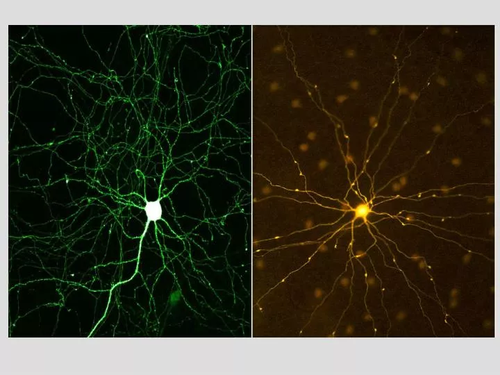
1 Synaptic terminals: Bring signals from other neurons. 3 Cell body: Integrates signals; Coordinates. 2 Dendrites: Receive signals from other neurons. 4 Action potential starts here. 5 Axon: Conducts the action potential. 6 Synaptic terminals: Transmit signals to other neurons. 7 Dendrites (of other neurons). Neurons Specialized “excitable” cells: receive input, integrate, send output synaptic terminal dendrite synapse
What parts of neurons are OUTSIDE the CNS? • All sensory, motor, and interneurons neurons • Sensory neuron dendrites & cell bodies AND motor neuron axons • Interneurons only • Motor neuron dendrites and interneuron axons
Neurons Neurons are electrical: • At rest, neurons maintain an electrical difference across their membrane • (-) inside cell; (+) outside cell • charge = about -70 mV Na+ pumped out, K+ pumped in, but K+ can leak out
Neurons Synapse: Region connecting two neurons or neuron and muscle synaptic terminal synaptic vesicle Neurotransmitter signals next neuron. Signal reaches end of axon. Synaptic vesicles release neurotransmitter. gap Neurotransmitter may excite or inhibit the next neuron neurotransmitter Receptor binds neurotransmitter. dendrite of postsynaptic neuron
(+) inside cell; (-) outside cell Na+ action potential (axon) (extracellular fluid) Na+ action potential (axon) (extracellular fluid) K+ Action Potentials Neurons Transmit Signals via Action Potentials: Action Potential (AP): The electrical signal passed along the length of a neuron • Neuron membrane polarized = charge difference • During action potential, Na+ channels open, flows in • Charge difference lost = depolarized • Triggers K+ channels to open, flows out • Repolarized
Action Potentials Na+ channels open K+ channels open Before K+ channels close, greater charge difference = hyperpolarized
Action Potentials Na+ and K+ channels are voltage gated – as first Na+ channels open, more triggered to open (i.e. positive feedback) Action potentials are propagated down the length of the neuron – after the AP, neuron resets itself (Na+ out, K+ in)
threshold resting potential potential (millivolts) EPSP IPSP time (milliseconds) Action Potentials Action potentials can be measured electrically: • Stimulation from a neighbor neuron excites the cell (brief increase in voltage = EPSP) • Inhibition from another neuron causes a brief decrease in voltage (IPSP)
Pre-synaptic terminal synaptic vesicle neurotransmitter potential (millivolts) EPSP dendrite of Post-synaptic neuron IPSP time (milliseconds) Action Potentials EPSP = excitatory post-synaptic potential (+) Neurotransmitter IPSP = inhibitory post-synaptic potential (-) Neurotransmitter Individual EPSP & IPSP weak
action potential potential (millivolts) threshold resting potential Less (-) More (-) time (milliseconds) Action Potentials • Sum of all excitatory & inhibitory ‘blips’ = summation • If threshold voltage is reached, an action potential occurs
Stimuli Input Information Coding in the Nervous System: • 1) Determine stimulus type (e.g. light / sound / touch) • All neurons use same basic signal • Wiring pattern in brain distinguishes stimuli • 2) Signal intensity of stimulus • All signals similar in size (all-or-none response) • Intensity coded by: • 1) Frequency of action potentials 2) # of neurons responding
1 fires slowly fires rapidly 1 2 silent 2 fires slowly 1 fires moderately 2 silent Stimuli Input Information Coding in the Nervous System:
Stimuli Input Information Coding in the Nervous System: • Integrate/coordinate signals • 4) Determine Output Neural Pathways Direct Behavior: • 1) Receptor: Detects stimulus • 2) Sensory neuron: Sends stimulus message • 3) Interneuron(s): Integrates stimuli • 4) Motor neuron: Activates effector • 5) Effectors: Performs function (muscle / gland)
Nervous System Organization Spinal Cord: • Myelin = Insulation around axons • Increases AP conduction rate
Nervous System Organization Spinal Cord:
What part of the spinal cord contains motorneuron cell bodies? • White matter • Dorsal root ganglia • Gray matter • Ventral roots
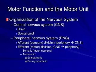
Nervous System Organization CNS Brain and Spinal Cord Motor Pathways Sensory Pathways Sensory neurons registering external stimuli Sensory neurons registering external stimuli PNS Somatic nervous system (voluntary) Autonomic nervous system (involuntary) Sympathetic nervous system "fight or flight" Parasympathetic nervous system "rest and repose"
If you are surprised by a “predator”, what happens in your nervous system? • Sympathetic nervous system increases heart rate • Somatic nervous system increases heart rate • Autonomic nervous system increases saliva • Parasympathetic NS increases saliva secretion
meninges skull hindbrain cerebellum pons medulla spinal cord Human Brain The Brain: • 1) Hindbrain: Automatic Behaviors • A) Medulla: Controls breathing, heart rate, blood pressure • B) Pons: Controls wake/sleep transitions; sleep stages • C) Cerebellum: Coordinates movement
pituitary gland pineal gland midbrain - Reticular Formation Human Brain The Brain: • 2) Midbrain: Relay / “Screening” Center • A) Reticular Formation: Controls arousal of brain • Filters sensory input from body • B) Visual / Auditory Reflex Centers
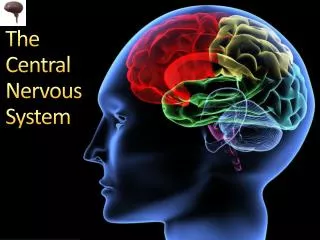
cerebral cortex corpus callosum Human Brain The Brain: • 3) Forebrain (Cerebrum): “Seat of Consciousness” • A) Cerebral Cortex • Two hemispheres (Connection = Corpus Callosum) • Left hemisphere controls right side of body (and vise versa)
Human Brain The Brain: Parietal Lobe Frontal Lobe Occipital Lobe Temporal Lobe • Forebrain (Cerebrum) • A) Cerebral Cortex • Four regions: • 1) Frontal: Primary motor area; complex reasoning • 2) Parietal: Primary sensory area • 3) Temporal: Primary auditory and olfactory areas • 4) Occipital: Primary visual area
Human Brain primary sensory area Frontal Lobe Parietal Lobe primary motor area leg premotor area trunk sensory association area arm higher intellectual functions hand Occipital Lobe face visual association area tongue speech motor area primary visual area primary auditory area auditory association area: language comprehension memory Temporal Lobe
Human Brain Motor and Sensory areas
Which is a correct match between brain region and function: • Parietal lobe : visual processing • Reticular formation : filters sensory input • Cerebellum : controls sleep stages • Cerebrum : dictates breathing rate
Hearing Words Seeing Words Reading Words Generating Verbs 0 max Human Brain Cortical Regions Involved in Different Tasks:
hypothalamus thalamus Human Brain The Brain: 3) Forebrain (Cerebrum) • B) Limbic System • Produce emotions; form memories • C) Thalamus • Relays information from body to limbic system / cerebral cortex
cerebral cortex limbic region of cortex corpus callosum thalamus hippocampus hypothalamus amygdala Human Brain The Brain: • B) Limbic System • Hypothalamus: Homeostatic control center • Amygdala: Produces sensations of pleasure, fear, or sexual arousal • Hippocampus: Formation of long-term memory • C) Thalamus • Relays to limbic system / cerebral cortex
Damage to the hippocampus could result in: • Failure to understand speech • Reduced fear response • Lack of homeostatic control • Loss of long-term memory formation
Things To Do After Lecture 14… • Reading and Preparation: • Re-read today’s lecture, highlight all vocabulary you do not understand, and look up terms. • Self-Quiz: Ch. 48 #1-3, 5; Ch. 49 #1, 2, 4, 5, 6 (correct answers in back of book) • Read chapters 48 & 49, focus on material covered in lecture (terms, concepts, and figures!) • Skim next lecture. • “HOMEWORK” (NOT COLLECTED – but things to think about for studying): • Explain the difference between the somatic and autonomic nervous systems. • Diagram a basic neuron – for sensory, motor, and interneurons explain the location of each region with respect to peripheral or central nervous system. • Compare and contrast the embryonic vertebrate brain with that of adults. • List the regions of the brain (with functions) from the “outside” of the brain, inward.