Download
1 / 50
500 likes | 548 Views
Cell Injury, Cell Death, and Adaptations. Cell adaptation. Hypertrophy Hyperplasia Atrophy Metaplasia. Hypertrophy Vs. Hyperplasia.

E N D
Cell adaptation Hypertrophy Hyperplasia Atrophy Metaplasia
B, Small spindle-shaped uterine smooth muscle cells from a normal uterus. Compare this with (C) large, plump hypertrophied smooth muscle cells from a gravid uterus.
Physiologic hypertrophy of the uterus during pregnancy. A, Gross appearance of a normal uterus (right) and a gravid uterus (left)
This is cardiac hypertrophy involving the left ventricle. The number of myocardial fibers does not increase, but their size can increase in response to an increased workload, leading to the marked thickening of the left ventricle in this patient with systemic hypertension.
Here is one of the nodules of hyperplastic prostate, with many glands along with some intervening stroma. The cells making up the glands are normal in appearance, but there are just too many of them.
The prominent folds of endometrium in this uterus opened to reveal the endometrial cavity are an example of hyperplasia. Cells forming both the endometrial glands and the stroma have increased in number. As a result, the size of the endometrium has increased. This increase is physiologic with a normal menstrual cycle.
The testis at the right has undergone atrophy and is much smaller than the normal testis at the left.
This is cerebral atrophy in a patient with Alzheimer disease. The gyri are narrowed and the intervening sulci widened.
Metaplasia of normal columnar (left) to squamous epithelium (right) in a bronchus, shown (A) schematically and (B) histologically.
Metaplasia of laryngeal respiratory epithelium has occurred here in a smoker. The chronic irritation has led to an exchanging of one type of epithelium (the normal respiratory epithelium at the right) for another (the more resilient squamous epithelium at the left).
Metaplasia of the normal esophageal squamous mucosa has occurred here, with the appearance of gastric type columnar mucosa.
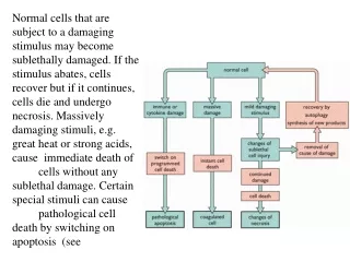
This microscopic appearance of myocardium is a mess because so many cells have died that the tissue is not recognizable. Many nuclei have become pyknotic (shrunken and dark) and have then undergone karorrhexis (fragmentation) and karyolysis (dissolution). The cytoplasm and cell borders are not recognizable.
Here is myocardium in which the cells are dying. The nuclei of the myocardial fibers are being lost. The cytoplasm is losing its structure, because no well-defined cross-striations are seen.
In this example, liver cells are dying individually (arrows) from injury by viral hepatitis. The cells are pink and without nuclei.
In this fetal thymus there is involution of thymic lymphocytes by the mechanism of apoptosis. Individual cells fragment and are consumed by phagocytes to give the appearance of clear spaces filled with cellular debris.
Two large infarctions (areas of coagulative necrosis) are seen in this sectioned spleen. Since the etiology of coagulative necrosis is usually vascular with loss of blood supply, the infarct occurs in a vascular distribution. Thus, infarcts are often wedge-shaped with a base on the organ capsule.
This is an example of coagulative necrosis. This is the typical pattern with ischemia and infarction (loss of blood supply and resultant tissue anoxia). Here, there is a wedge-shaped pale area of coagulative necrosis (infarction) in the renal cortex of the kidney.
The contrast between normal adrenal cortex and the small pale infarct is good. The area just under the capsule is spared because of blood supply from capsular arterial branches. It illustrates the shape and appearance of an ischemic (pale) infarct well.
The liver shows a small abscess here filled with many neutrophils. This abscess is an example of localized liquefactive necrosis.
This is liquefactive necrosis in the brain in a patient who suffered a "stroke" with focal loss of blood supply to a portion of cerebrum. This type of infarction is marked by loss of neurons and neuroglial cells and the formation of a clear space at the center left.
Grossly, the cerebral infarction at the upper left here demonstrates liquefactive necrosis. Eventually, the removal of the dead tissue leaves behind a cavity.
This is fat necrosis of the pancreas. Appear grossly as the soft, chalky white areas seen here on the cut surfaces.
This is the gross appearance of caseous necrosis in a hilar lymph node infected with tuberculosis. The node has a cheesy tan to white appearance.
This is more extensive caseous necrosis, with confluent cheesy tan granulomas in the upper portion of this lung in a patient with tuberculosis.
Microscopically, caseous necrosis is characterized by acellular areas, as the tissue architecture is completely lost (at the upper right), surrounded by a granulomatous inflammatory process.
This is gangrene of the lower extremity. In this case the term "wet" gangrene is more applicable because of the liquefactive component from superimposed infection in addition to the coagulative necrosis from loss of blood supply. This patient had diabetes mellitus.
Here is fatty change of the liver due to accumulation of lipid in the cytoplasm of hepatocytes.
Here are Mallory bodies (the red globular material) composed of cytoskeletal filaments in liver cells chronically damaged from alcoholism. These are a type of "intermediate" filament between the size of actin (thin) and myosin (thick). (proteins accumulation)
The brown coarsely granular material in macrophages in this alveolus is hemosiderin that has accumulated as a result of the breakdown of RBC's and release of the iron in heme. The macrophages clear up this debris, which is eventually recycled.
Here is anthracotic pigment in macrophages in a hilar lymph node. Anthracosis is nothing more than accumulation of carbon pigment from breathing dirty air. Smokers have the most pronounced anthracosis.
This is dystrophic calcification in the wall of the stomach. At the far left is an artery with calcification in its wall. There are also irregular bluish-purple deposits of calcium in the submucosa.
Calcification of the aortic valve. A view looking down onto the unopened aortic valve in a heart with calcific aortic stenosis. The semilunar cusps are thickened and fibrotic. Behind each cusp are large, irregular masses of dystrophic calcification that will prevent normal opening of the cusps.