Download
1 / 40
500 likes | 776 Views
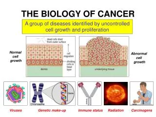
The Biology of Cancer. Peter Curtin, M.D. Professor of Clinical Medicine Clinical Director, Blood and Marrow Transplantation Division September 12, 2013. The Hallmarks of Cancer.

E N D
The Biology of Cancer Peter Curtin, M.D. Professor of Clinical Medicine Clinical Director, Blood and Marrow Transplantation Division September 12, 2013
The Hallmarks of Cancer • A conceptual framework for understanding the biological diversity of cancer and to understanding the multistep process of progression from normal cells to a neoplastic state • Hallmarks of Cancer are eight, acquired, functional capabilities that allow cancer cells to survive, proliferate and disseminate • First proposed by Hanahan and Weinberg in 2000 • Updated and expanded by Hanahan and Weinberg (Cell, 2011)
The Hallmarks of Cancer • 1. Sustaining proliferative signaling • 2. Evading growth suppressors • 3. Resisting cell death • 4. Enabling replicative immortality • 5. Inducing angiogenesis • 6. Activating invasion and metastasis • 7. Deregulating cellular energetics • 8. Avoiding immune destruction
1. Sustaining proliferative signaling • Cancer cells need to sustain chronic proliferation • Growth promoting signals are normally conveyed by growth factors binding to cell surface receptors which typically contain intracellular tyrosine kinase domains • The tyrosine kinase domain transmits signals via downstream signaling protein pathways to control progression through the cell cycle, cell growth and to influence cell survival and energy metabolism
Sustaining proliferative signaling • Mechanisms to sustain proliferative signaling: • 1. Cancer cells produce growth factors themselves (autocrine stimulation) • 2. Stimulate nearby normal (stromal) cells to produce growth factors • 3. Increased levels of cell surface receptor proteins • 4. Structurally abnormal receptors, active in the absence of growth factor • 5. Constitutive activation of signaling proteins downstream from the receptor
2. Evading growth suppressors • Cancer cells must overcome programs that negatively regulate cell proliferation • These programs depend on the action of tumor suppressor genes that normally govern the decision of cells to proliferate or to undergo program cell death (apoptosis) • Tumor suppressor genes have typically been discovered when inactivation leads to the development of cancer • The retinoblastoma (RB) protein and p53 protein are two tumor suppressors (among many described)
Evading growth suppressors • RB protein integrates extracellular and intracellular signals to decide whether the cells should proceed thru growth and division • Loss of RB function (by deletion or mutation) removes a gatekeeper of cell cycle progression resulting in persistent cell proliferation • p53 protein senses intracellular stress and abnormality • If DNA damage is present or if growth promoting signals, oxygen or glucose are suboptimal, p53 can stop cell cycle progression until these conditions normalize • If overwhelming or irreparable damage to intracellular systems occurs, p53 can trigger program cell death (apoptosis)
3. Resisting cell death • Programmed cell death by apoptosis is a natural barrier to the development of cancer • The apoptotic machinery is composed of upstream regulator proteins and downstream effector proteins • Regulators • Extracellular/extrinsic pathway: Fas ligand and receptor, Tumor necrosis factor (TNF)/TNF receptor • Intracellular/intrinsic pathway: senses intracellular signals • Effectors: inactive proteases (caspases 8 and 9) are activated initiating a proteolytic cascade leading to cellular disassembly and consumption (apoptosis)
Resisting cell death: The Bcl-2 family • The Bcl-2 family of pro- and anti-apoptotic regulatory proteins: • Bcl-2 (and relatives) are inhibitors of apoptosis, acting by binding to and inactivating two pro-apoptotic proteins, Bax and Bak, that live in the mitochondrial membrane • Bax and Bak, when released from Bcl-2 binding (and inhibition), disrupt the outer mitochondrial membrane, releasing cytochrome c which activates the cascade of proteolyticcapsases leading to the cellular changes of apoptosis • Bcl-2 interacts with Bax and Bak via BH3 interaction domains • Other proteins that sense cellular abnormalities contain BH3 domains (“BH3-only” proteins) can activate apoptosis by interfering with Bcl-2 or by activating Bax or Bak directly
Resisting cell death Sensors that trigger apoptosis: • DNA damage sensor functions via p53 • Insufficient survival factor signaling (e.g. IL-3 for lymphocytes) • Hyperactive signaling by some oncoproteins (e.g. Myc) • Each of these can activate BH3-only proteins to induce apoptotic cascade
Resisting cell death: • Mechanisms to limit or circumvent apoptosis: • Loss of the p53 tumor suppressor function • Increased expression of anti-apoptotic regulators (e.g. Bcl-2) • Increased expression of survival signals (e.g. IL-3) • Decreased expression of pro-apoptotic regulators (e.g. Bax and Bak) • Interrupting the extrinsic apoptotic pathway
4. Enabling replicative immortality • Normal cells pass thru a limited number of cell (division) cycles before becoming senescent (alive but non-proliferative) or undergoing crisis leading to cell death • In the culture, repeated cycles of cell division lead to senescence and then crisis, resulting in death of the majority of cells. • Rare cells that survive crisis exhibit unlimited replicative potential and are said to be immortalized (a characteristic of established cell lines) • Telomeres, multiple tandem copies of 6 nucleotide repeats protecting the ends of chromosomes, are central to limiting the number of division cycles that a normal cell can undergo and, conversely, to the unlimited proliferation capacity of malignant cells
Enabling replicative immortality:Telomeres and Telomerase • In normal cells, telomeres progressively shorten with each cell division • Eventually lose the ability to protect the ends of chromosomes from end-to-end fusions • End-to-end chromosomal fusion results in scrambling of the karyotype and leads to apoptosis • The length of telomeric DNA in a cell dictates how many cell generations its offspring can pass through prior to telomere erosion and cell death • Telomerase is a specialized DNA polymerase that adds telomere repeats to the ends of telomeres • Telomerase is almost absent in normal cells but is present in immortalized cells
Enabling replicative immortality:Telomeres and Telomerase • Mechanisms to achieve replicative immortality: • Increase expression of telomerase • Activation of an alternative, recombination based telomere-maintenance mechanism (less common)
5. Inducing angiogenesis • Tumors require new blood vessel formation to survive and grow • In adult life, normal angiogenesis is required as a part of wound healing and during the female reproductive cycle • During tumor development and progression the “angiogenic switch” is activated and remains on to support crowding of new vessels stained neoplastic growth • The angiogenic switch is controlled by factors that either induce or oppose angiogenesis • Vascular endothelial growth factor (VEGF) signals thru receptor tyrosine kinases (VEGFR) to promote angiogenesis • Fibroblast growth factor (FGF) also is pro-angiogenic
Inducing angiogenesis • Thrombospondin-1 (TSP-1) and fragments of Plasmin (angiostatin) and type 18 collagen (endostatin)are endogenousinhibitors of angiogenesis • Theseproteinsserve as regulators of normal transitoryangiogenesisduringhealing and mayalsoact as barrierstocancerdrivenangiogenesis
Inducing angiogenesis • Mechanisms to induce angiogenesis: • Increased VEGF expression due to hypoxia or oncogene signaling (Ras and Myc) • Increased expression of FGF and other pro-angiogenic molecules • Leukocytes (macrophages, neutrophils, mast cells) infiltrating pre-malignant and malignant lesions can activate the angiogenic switch and sustain ongoing angiogenesis
6. Activating invasion and metastasis • Carcinomas developed alterations in shape as well as in their attachment to other cells and to the extracellular matrix • E-cadherin is a cell-to-cell adhesion molecule that helps to assemble epithelial cells into sheets and to maintain quiescence • E-cadherin is frequently down regulated and occasionally inactivated by mutation in carcinomas • N-cadherin is normally expressed in migrating neurons and mesenchymal cells during organogenesis • N-cadherin is often upregulated in invasive carcinoma cells
Activating invasion and metastasis • The invasion-metastasis cascade: • Local invasion followed by intravasation of cancer cells into nearby blood and lymph vessels • Transit via lymph and blood followed by escape from vessels into distant parenchyma (extravasation) • Formation of small tumor nodules (micrometastases) • Growth into macroscopic tumors (colonization)
Activating invasion and metastasis • “Epithelial-mesenchymal transition” (EMT) is a developmental regulatory program involved in embryonic morphogenesis and wound healing that epithelial cancers co-opt to acquired the ability to invade, resist apoptosis and disseminate • EMT and related migratory processes in embryogenesis are controlled by a set of transcriptional factors (Snail, Slug, Twist etc.) • These factors are expressed widely in cancer and are important in invasion and metastasis • The factors induce loss of adherans junctions, conversion from round to spindle shape, expression of matrix-degrading enzymes, increased motility and resistance to apoptosis • The EMT program in embryogenesis and cancer is influenced by signals from neighboring stromal cells
Activating invasion and metastasis • Crosstalk between cancer cells and nearby stromal cells is involved in invasive growth and metastasis: • Mesenchymal stem cells produce CCL5 in response to signals from cancer cells; CCL5 acts on the cancer cells to stimulate invasion • Cancer cells produce IL-4 to activate macrophages to elaborate matrix-degrading enzymes to facilitate invasion • Tumor associated macrophages supply epidermal growth factor to breast cancer cells; the cancer cells release CSF-1 to stimulate the macrophages • Colonization (growth into macro-metastases) requires adaptation to a new environment
7. Deregulating cellular energetics • Chronic cell proliferation requires adjustment of energy metabolism • Normal cells use glucose via glycolysis under anaerobic conditions but favor oxidative phosphorylation under aerobic conditions • Cancer cells can reprogram glucose metabolism to favor glycolysis even under aerobic conditions • Glycolysis is much less efficient than oxidative phosphorylation • Cancer cells compensate by up-regulating glucose transporters (GLUT1) to increase glucose uptake into the cell • Use of glycolysis is associated with activated oncogenes (RAS, MYC), with mutant tumor suppressors (p53) and can be further increased in the setting of hypoxia, present in many tumors
Deregulating cellular energetics • Why do cancer cells favor glycolysis? • Perhaps diversion of glycolytic intermediates into biosynthetic pathways generating nucleosides and amino acids facilitates biosynthesis of macromolecules required for proliferation • Similar metabolic changes are found in rapidly dividing embryonic tissues suggesting a role in supporting active cell proliferation • Some tumors have two populations of cells, one more hypoxic that use glycolysis and secrete lactate, another, better oxygenated that imports lactate and uses it as an energy source in oxidative phosphorylation • Oxygenation in tumors fluctuates, both in time and space, due to the instability and disorganization of tumor vasculature
8. Avoiding immune destruction • Theory of Immune Surveillance: • Suggests that the immune system recognizes and eliminates the vast majority of incipient cancer cells • Tumors that develop either manage to avoid immune detection or limit immunologic killing • Increases in certain cancers in immunocompromised individuals supports this theory; however, most of these cancers are virally induced, unlike the majority of cancers • Mice engineered to lack cytotoxic T lymphocytes (CTLs), helper T cells or natural killer (NK) cells all developed more carcinogen-induced tumors and more rapidly growing tumors • Supports the role of cellular immunity in tumor eradication
Avoiding immune destruction • Transplant experiments showed that tumors arising in immunodeficient mice can only be successfully transplanted into immunodeficient mice while tumors arising in immunocompetent mice can be successfully transplanted into both types of mice • Suggests that highly immunogenic cancers are eliminated in immunocompetent mice (“immunoediting”), leaving only weakly immunogenic cancers to survive and be transplanted into both types of mice • In immunodeficient mice, highly immunogenic cancers survive and can be transplanted into immunodeficient mice but will not survive in immunocompetent mice
Avoiding immune destruction • Clinical observations supporting antitumor immune response: • Patients with colon and ovarian tumors that are heavily infiltrated with CTLs and NK cells have a better prognosis • Immunosupressed organ transplant recipients have developed donor-derived cancers, suggesting that in the tumor-free donor, the cancer was held in check by an intact immune system • Highly immunogenic tumors may have ways of disabling components of the immune system by secreting TGF-beta or other immunosuppresive factors or by recruiting regulatory T cells (T regs) or myeloid-derived supressor cells which can both supress the function of cytotoxic T lymphocytes
Two Enabling Characteristics of Cancer • Genome Instability and Mutation • Tumor Promoting Inflammation
Genome Instability and Mutation • Acquisition of Hallmarks depends on a succession of alterations in the genomes of neoplastic cells • Certain mutations offer a selective advantage leading to outgrowth of the dominant clone • Multistep tumor progression is a series of clonal expansions each triggered by the chance acquisition of an enabling mutation • Cancer cells increase mutation rate by: increased sensitivity to mutagenic agents thru breakdown in the genomic maintenance machinery • Mutation rate also increased by compromising the systems that monitor genomic integrity and force damaged cells into senescence or apoptosis
Tumor-Promoting Inflammation • Tumor cells are typically infiltrated by cells of the immune system • Immune cells aiming to eradicate the tumor vs. enhancing tumor development and progression • Inflammatory cells can supply growth factors to sustain proliferative signaling, survival factors limiting cell death, pro-angiogenic factors, matrix-modifying enzymes that facilitate angiogenesis, invasion and metastasis and induction signals leading to activation of EMT • Inflammation sometimes demonstrable at early stages of tumor development and may foster development from pre-malignant lesion to cancer • Inflammatory cells can release chemicals, particularly reactive oxygen species that are mutagenic, hastening malignant progression • Role of the tumor microenviroment in cancer
The Cancer Stem Cell • Cancers are comprised of heterogenous populations of cells comprised of regions with various degrees of differentiation, proliferation, vascularity, inflammation and invasiveness • Cancer stem cells represent a further degree of cellular heterogeneity and are likely a common constituent of most tumors • Defined by their ability to efficiently seed new tumors in recipient host mice • Initially identified in hematologic malignancies but subsequently found in solid tumors as well • Cell surface markers and gene transcription profiles similar to those of normal tissue stem cells • Cancer stems cells are likely to be particularly resistant to conventional chemotherapy and may require novel therapeutic approaches
The Cancer Genome Era • The project to determine the sequence of an entire human genome began in 1990 and was completed in 2003 • Several large sequencing labs in the US and abroad participated • The cost of the Human Genome Project was $2.7 billion • Subsequent improvements in technology has allow faster and cheaper sequencing • Currently genome sequencing may take several weeks and cost $10,000 or less • Identify important genetic mutations that are biologic drivers for given cancers or subsets of cancers, improve prognostication, provide additional targets for drug development, provide foundation for cancer therapy that is tailored to each cancer