Download
1 / 19
190 likes | 196 Views
Internal medicine L-4 Liver cirrhosis & portal hypertension. Prepared by: Kholod Hamad MSc clinical pharmacy, BCPS. Cirrhosis.

E N D
Internal medicineL-4Liver cirrhosis & portal hypertension Prepared by: Kholod Hamad MSc clinical pharmacy, BCPS
Cirrhosis • Cirrhosis is defined as fibrosis of the hepatic parenchyma resulting in nodule formation, altered hepatic function, restricted venous outflow, and portal hypertension. • It results from a sustained wound-healing response to chronic or acute liver injury from a variety of causes, the most common being chronic viral hepatitis chronic alcohol consumption.
The illustration below shows the nodular changes that occur in cirrhosis
Cirrhosis Hepatocytes fibrous tissue Complications • Portal hypertension • Varices and ascites • Hepatic encephalopathy (HE) • Coagulopathy
Child-Pugh Classification of the Severity of Cirrhosis Class A = total score of 5 or 6 Class B = total score of 7–9 Class C = total score of 10 or more
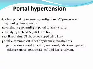
GastroesophagealVarices Background • a. Resistance to blood flow within the liver secondary to cirrhosis results in the development of portal hypertension. Collateral blood vessels (e.g., esophageal varices) are formed because of this increased resistance to blood flow. • b. Variceal hemorrhage may occur in around 25%–35% of patients with cirrhosis and varices; mortality rates are as high as 30%–50% per bleed; recurrence rates are as high as 70% within the first 6 months after an initial bleed.
Management of acute variceal bleeding • Fluid resuscitation and hemodynamic stabilization • Endoscopy to assess the extent of disease with potential intervention • Medical management of acute variceal bleeding to reduce splanchnic blood flow and portal pressure (Vasopressin, Octreotide) • Antibiotic therapy
Ascites • Definition: Free fluid in the abdominal cavity secondary to increased resistance within the liver (forces lymphatic drainage into the abdominal cavity) and reduced osmotic pressure within the bloodstream (hypoalbuminemia); develops at a 5-year cumulative rate of 30% in compensated liver disease
Clinical features: Protuberant abdomen, shifting dullness, fluid wave, bulging flanks, abdominal pain • Diagnosis a. Clinical features b. Abdominal ultrasonography c. Paracentesis. Can use serum-ascites albumin gradient, calculated by subtracting the ascites albumin concentration from the serum albumin concentration; a value greater than 1.1 indicates ascites secondary to portal hypertension
Treatment of Ascites • Alcohol cessation if alcohol induced • Dietary sodium restriction and water restriction • Diuretics: combination of furosemide and spironolactone (ratio 40:100) • If tense ascites is present, may use large-volume paracentesis. Administer albumin at a dose of 6–8 g/L of ascitic fluid removed over 5 L • If refractory ascites is present, may consider midodrine add-on therapy
Spontaneous Bacterial Peritonitis Background • Infection of previously sterile ascitic fluid without an apparent intra-abdominal source. SBP is considered a primary, as opposed to secondary, peritonitis. • May be present in 10%–30% of hospitalized patients with cirrhosis and ascites c. A ssociated with 20%–40% of in-hospital mortality; poor prognosis after recovery, with 2-year survival after initial episode reported as about 30%
Pathophysiology • Principal theory is seeding of the ascitic fluid from an episode of bacteremia. • The bacteria present are usually enteric pathogens; thus, they may enter the blood because of increases in gut permeability secondary to portal hypertension, suppression of hepatic reticuloendothelial cells, or translocation of the gut wall and dissemination through the mesenteric lymph system.
Reduced opsonic activity of the ascitic fluid and alterations in neutrophil function may also be contributing factors. • Enteric gram-negative pathogens are most commonly involved, and more than 90% of cases involve a single bacterial species.
Management • Broad-spectrum antibiotictherapyto coverEscherichia coli, Klebsiellapneumoniae, and Streptococcus pneumoniae. • IV Cefotaxime or Oral ofloxacin • Antibiotic prophylaxiswithdailynorfloxacin400mgordouble-strengthtrimethoprim-sulfamethoxazole.
References • Clinical pharmacy & therapeutics, Edi 5, chapter 16 Liver disease. Page 238