Download
1 / 2
20 likes | 129 Views
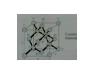
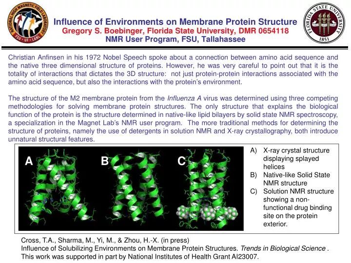
A. B. C. Influence of Environments on Membrane Protein Structure Gregory S. Boebinger , Florida State University, DMR 0654118 NMR User Program, FSU, Tallahassee.

E N D
A B C Influence of Environments on Membrane Protein StructureGregory S. Boebinger, Florida State University, DMR 0654118NMR User Program, FSU, Tallahassee Christian Anfinsen in his 1972 Nobel Speech spoke about a connection between amino acid sequence and the native three dimensional structure of proteins. However, he was very careful to point out that it is the totality of interactions that dictates the 3D structure: not just protein-protein interactions associated with the amino acid sequence, but also the interactions with the protein’s environment. The structure of the M2 membrane protein from the Influenza A virus was determined using three competing methodologies for solving membrane protein structures. The only structure that explains the biological function of the protein is the structure determined in native-like lipid bilayers by solid state NMR spectroscopy, a specialization in the Magnet Lab’s NMR user program. The more traditional methods for determining the structure of proteins, namely the use of detergents in solution NMR and X-ray crystallography, both introduce unnatural structural features. X-ray crystal structure displaying splayed helices Native-like Solid State NMR structure Solution NMR structure showing a non-functional drug binding site on the protein exterior. Cross, T.A., Sharma, M., Yi, M., & Zhou, H.-X. (in press) Influence of Solubilizing Environments on Membrane Protein Structures. Trends in Biological Science . This work was supported in part by National Institutes of Health Grant AI23007.
A B Influence of Environments on Membrane Protein Structure Gregory S. Boebinger, Florida State University, DMR 0654118NMR User Program, FSU, Tallahassee Despite Christian Anfinsen’s acknowledgement in 1972 that protein interactions with their environments are important, it has long been erroneously thought that the amino acid sequence alone was responsible for the three dimensional structure of proteins. This error likely results from the fact that more than 99% of the protein structures determined to date are of water soluble proteins in a uniform aqueous environment. However, high-magnetic-field NMR is finding that the complex and heterogeneous environment of membranes directly impacts the structure of membrane proteins, particularly the structures of those proteins that span the cellular membrane. Since native membranes are too complex for current NMR technology, it is essential that the membrane environment be adequately mimicked when determining the structure of membrane proteins. These results show that a synthetic lipid bilayer provides a much more accurate environment for membrane proteins than the detergents typically used for solution NMR and X-ray crystallography. In the X-ray structure of M2 protein the detergent molecules are wedged in three of the four helical interfaces causing the helices to splay apart. The detergent micelle permits a thicker hydrophobic environment than the native membrane and as a result the helices pack more parallel to each other preventing the drug from binding in the pore between the four helices in this solution NMR structure.