Download
1 / 113
1.17k likes | 1.85k Views
ABNORMALITIES OF TEETH. 牙齒的異常 - 環境與發育的影響. Environmental Alterations of Teeth Developmental Alterations of Teeth. 王文岑 高雄醫學大學 牙醫學系 wcwang@kmu.edu.tw . ENVIRONMENTAL ALTERATIONS OF TEETH. Developmental tooth defects Turner’s tooth Hypoplasia caused by antineoplastic therapy

E N D
ABNORMALITIES OF TEETH 牙齒的異常-環境與發育的影響 Environmental Alterations of Teeth Developmental Alterations of Teeth 王文岑 高雄醫學大學 牙醫學系 wcwang@kmu.edu.tw
ENVIRONMENTAL ALTERATIONS OF TEETH • Developmental tooth defects • Turner’s tooth • Hypoplasia caused by antineoplastic therapy • Fluorosis • Syphilitic hypoplasia • Postdevelopmental structure loss • Tooth wear • Internal and external resorption • Discolorations of teeth • Intrinsic stains • Extrinsic stains • Localized disturbances in eruption • Primary impaction • Ankylosis
Enamel development • Three stages:1. Matrix formation: protein laid down 2. Mineralization: minerals deposition, majority of original prot. removed-- diffuse, opaque white, soft enamel 3. Maturation: final mineralization-- translucent, hard enamel • Amelogenesis imperfecta • Enamel hypoplasia
Enamel development • No remodeling after initial formation • Timing of ameloblastic damage has a great impact on location & appearance of the defect • Development of crown : from 14th week of gestation to 12 months of age in deciduous dentition; 6 months to 15 y/o in permanent dentition • Neonatal ring on deciduous enamel and deposition with a rate of 0.023mm/day
See Box 2-2 Factors associated with enamel defects Systemic- 1. Birth-related trauma: premature birth 2. Chemicals: antineoplastic C/T, fluoride, tetracycline 3. Chromosomal abnormalities: trisomy 21 4. Infections: chicken pox, CMV, syphilis 5. Inherited diseases: Vit.D-dependent rickets 6. Malnutrition: Vit. A deficiency 7. Metabolic disorders: hypoparathyroidism, maternal diabetes 8. Neurologic disorders: cerebral palsy
See Box 2-2 Factors associated with enamel defects Local- 1.Local acute mechanical trauma 2. Electric burn 3. Irradiation 4. Local infection: periapical inflammatory disease
Clinical and Radiographic Features Environmental enamel defects: 1.Hypoplasia:pits, grooves or large area of missing enamel 2. Diffuse opacities: variation in translucency, normal thickness, white opacity without clear boundary 3. Demarcated opacities: increased opacity, a sharp boundary with adjacent normal enamel, normal thickness
Turner’s hypoplasia, Turner’s tooth • Permanent teeth • Periapical inflammatory disease of the overlying deciduous tooth, less frequently in anterior teeth • Traumatic injury- not rare -45% children sustain injury to their deciduous teeth, 23% permanent teeth development disturbed Turner’s hypoplasia secondary to previous trauma
Hypoplasia caused by antineoplastic therapy • Under 12 y/o, esp. under 5y/o • Age at treatment, forms of therapy Chemotherapy- • Less alteration than radiation • Increased number of enamel hypoplasia and discolorations, slight smaller tooth size, radicular hypoplasia
Radiotherapy- • 0.72 Gy related to mild defects in enamel, dentin (一般成人頭頸癌照射一次約為2Gy) • Dose, radiation field
Developmental radicular hypoplasia and microdontia caused by radiotherapy
Hypodontia, microdontia, radicular hypoplasia, enamel hypoplasia, mandibular hypoplpasia, reduced in vertical development of lower 1/3 of face • Mandibular hypoplpasia may caused by Radiation →impaired root development →reduced alveolar bone growth • Cranial radiation→ altered pituitary gland function→ growth failed
*Dental fluorosis • 1901, Dr. Frederick S. McKay: Colorado brown stain • 1909, Dr. F.L. Robertson in Bauxite, Arkansas • 1930, H.V. Churchill: high concentration of fluoride of Bauxite(13.7ppm) and Colorado • 1931, Dr. H. Trendley Dean: association between fluoride, dental fluorosis and prevalence of caries among children • 1.0 ppm reduced caries by 50~70% and associated with low and mild mottled enamel • 0.7~1.2 ppm water fluoridation was recommended after 1962, currently 0.7ppm is recommended due to increased dental fluorosis
Dental fluorosis • Retention of the amelogeninprotein in enamel structure→ hypomineralized enamel→ permanent hypomaturation→ increased surface and subsurface porosity→ alters light reflection and create white, chalky area
Dental fluorosis • Critical period for clinical dental fluorosis is the 2nd and 3rd year of life, dose dependent • Caries resistant
Syphilitic hypoplasia • Congenital syphilis • Hutchinson’s incisors & mulberry molars
POSTDEVELOPMENTAL LOSS OF TOOTH STRUCTURE • Begin from enamel surface (tooth wear): Attrition, abrasion, erosion, abfraction • Begin from dentin, cemental surface: internal or external resorption
Attrition • Tooth to tooth contact during occlusion and mastication, some are physiologic • Accelerated by: poor quality or absent enamel, premature contact, intraoral abrasives, erosion, grinding habits • Incisal, occlusal and interproximal surfaces
Abrasion • Pathologic loss of tooth structure or restoration secondary to the action of an external agent (ex. Toothbrush, hair grips, toothpicks, chewing tobacco, biting thread, dental flossing…) • Toothbrush abrasion: horizontal buccal cervical notches of exposed radicular cementum and dentin with smooth surface. • Greater on prominent teeth ( canines, premolars , and teeth adjacent to edentulous area) and side of the arch opposite to the dominant hand • Demastication- when tooth wear is accelerated by chewing an abrasive substance between opposing teeth (both attrition and abrasion)
Improper use of hair grips Abrasion Long-term use of tobacco pipe
Erosion • Chemical process, exposure to acidic foods or drinks, medications (chewable Vit. C, aspirin), involuntary regurgitation (ex. esophagitis, pregnancy), voluntary regurgitation (ex. psychologic problems, bulimia) • Perimolysis- dental erosion from gastric secretion • Facial surface of maxillary anteriors affected-dietary source • Posterior teeth extensive loss of occlusal surface, and palatal surface concave dentin surrounded by an elevated enamel rim- regurgitation of gastric secretion
Erosion concave dentin surrounded by an elevated enamel rim
Erosion A bulimia patient
Abfraction • Repeated tooth flexure caused by occlusal stresses (tensile stress) → concentrate at the cervical fulcrum → may produce disruption in the chemical bonds of enamel crystal →cracked enamel can be lost or removed by erosion or abrasion • Wedge-shaped cervical defects, deep, narrow V-shaped, not allow toothbrush to contact base; if the defect, often affect a single tooth • Almost exclusively on facial surface and more often in bruxism, higher in mandibular dentition
Treatment and prognosis of tooth wear • Resolve pain and sensitivity • Identify the cause of tooth structure loss • Protection
INTERNAL & EXTERNAL RESORPTION • Internal resorption- by cells located in pulp, rare • Follows injury to pulp tissues, physical trauma or caries, continue as long as vital pulp remains, may result in communication of the pulp and PDL • External resorption-by cells in PDL, common
Clinical and Radiographic Features Internal resorption- • Inflammatory resorption- dentin replaced by inflamed granulation tissue • Pink tooth of Mummery: internal resorption involved coronal pulp Balloonlike enlargement of the canal • Replacement, or metaplastic absorption- pulpal dentinal walls are replaced by bone or cementum-like bone
Clinical and Radiographic Features External resorption- • Moth-eaten loss of tooth structure, less well-defined and variation in density in radiography • Most involved apical or midportions of root, occasionally, begin from cervical (invasive cervical resorption)
Histopathologic Feature • Increased cellularity, vascularity and collagenization • Numerous multinucleated dentinoclasts • Inflammatory cells infiltration
Treatment and prognosis • Internal resorption- • Removal of all soft tissue from site of resorption • Endodontic treatment before perforation in internal resorption • Placement of calcium hydroxide paste for remineralization • Surgical exposure and restoration • Extraction • External resorption- • Identification and elimination the accelerating factor
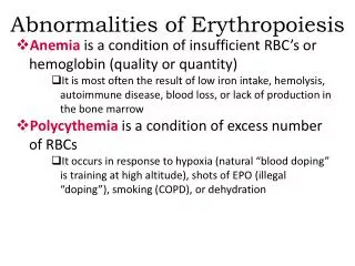
ENVIRONMENTAL DISCOLORATION OF TEETH • Extrinsic- surface accumulation of exogenous pigment • Intrinsic-secondary to endogenous factors that result in discoloration of underlying dentin
Extrinsic stains • Bacterial- Chromogenic bacteria, green, black-brown, orange coloration Frequently in children, labial surface of maxillary ant. in gingival third • Iron- formation of ferric sulfide • Tobacco • Food and beverage- chlorophyll • Gingival hemorrhage- Hb. breakdown to biliverdin • Restorative material – ex. Amalgam • Medications- iron, iodine, silver nitrate, chlorhexidine, stannous fluoride
Intrinsic stains • Amelogenesis imperfecta • Dentinogenesis imperfecta • Dental fluorosis • Erythropoietic porphyria – • autosomatic recessive disorder of porphyrin metabolism, increased synthesis and excretion of porphyrins and their related precursors • Porphyrin deposition in teeth, reddish-brown coloration, red fluorescence when exposed to a Wood’s UV light • Present both in dentin and enamel in deciduous teeth, but only dentin affected in permanent teeth
Erythropoietic porphyria Hyperbilirubinemia
Intrinsic stains • Hyperbilirubinemia- bilirubin, breakdown product of RBC, jaundance (yellow-green discoloration), erythroblastosis fetalis, biliary atresia • Biliverdin deposition, green discoloration of teeth (chlorodontia) • Ochronosis-alkaptonuria, blue-black discoloration • Trauma- coronal discoloration, pulp necrosis • Localized RBC breakdown
Intrinsic stains • Medications- Tetracycline (bright yellow to dark brown), chlortetracycline (gray-brown), oxytetracycline (yellow) , minocycline hydrochloride • Time of administration dose, duration • Avoid from pregnancy up to 8 yrs of age
Minocycline hydrochloride • Tx for Acne • Blue-gray from incisal 3/4, to dark green or black in roots, also affect developed teeth • Skin, nail, sclera, conjunctiva, thyroid, bone discoloration in susceptible individuals Stained alveolar bone
Treatment and prognosis • Extrinsic stains- polishing • Intrinsic stains- bleaching, bonded restoration, crowns
LOCALIZED DISTURBANCES IN ERUPTION • PRIMARY IMPACTION-Teeth cease to eruption before emergence • ANKYLOSIS-Cease of eruption after emergence and anatomicfusion of tooth cementum or dentin with alveolar bone
Impaction • 3rd molars, maxillary canines, mandibular premolars, mandibular canines, maxillary premolars, maxillary central incisors, maxillary lateral incisors, and mandibular second molars; usually angulated or diverted Factors associated with impaction: • Crowding and deficient maxillofacial development • Overlying cysts or tumors • Trauma • Reconstructive surgery • Thickened overlying bone or soft tissue • A host of systemic disorders, diseases or syndromes
Classification : • Partially erupted or full bony impaction • according to angulation: Mesioangular, distoangular, vertical, horizontal or inverted • Eruption sequestrum
Treatment and Prognosis Choice of treatment: • Long-term observation • Orthodontically assisted eruption • Transplantation • Surgical removal The risks associated with nonintervention: • Crowding dentition • Resorption and worsening of the periodontal status of adjacent teeth • Development of pathologic conditions, ex infections, cysts or tumors
The risks associated with intervention: • Transient or permanent sensory loss • Alveolitis • Trismus • Infection • Fracture • TMJ injury • Periodontal injury • Injury to adjacent teeth
ANKYLOSIS Infraocclusion, secondary retention, submergence, reimpaction, reinclusion
ANKYLOSIS Clinical And Radiographic Features • Pathogenesis is unknown, may be secondary to many factors and result in PDL barrier deficiency. • May occur at any age, any tooth • Most affect 8~9yr-old children and D , E , D , E • PDL absent • Occlusal, periodontal problems, impaction of the underlying teeth Treatment and Prognosis • Variable: extraction, orthodontics, segmental osteotomy
SHAPEGemination, Fusion, Concrescence Accessary cusps Dense in dente Ectopic Enamel Taurodontism Dilaceration Hypercementosis Supernumerary roots NUMBER Hypodontia Hyperdontia SIZE Microdontia Macrodontia STRUCTURE Amelogenesis imperfecta Dentinogenesis imperfecta Dentin dysplasia I & II Regional odontodysplasia DEVELOPMENTAL ALTERATIONS OF TEETH