Download
1 / 55
790 likes | 1.74k Views
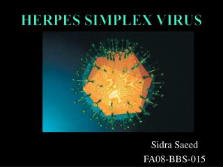
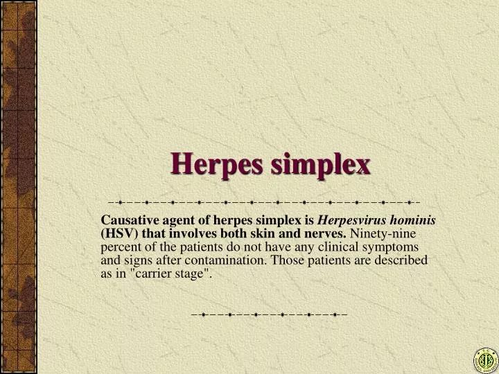
Herpes simplex. Causative agent of herpes simplex is Herpesvirus hominis (HSV) that involves both skin and nerves. Ninety-nine percent of the patients do not have any clinical symptoms and signs after contamination. Those patients are described as in "carrier stage".

E N D
Herpes simplex Causative agent of herpes simplex is Herpesvirus hominis (HSV) that involves both skin and nerves. Ninety-nine percent of the patients do not have any clinical symptoms and signs after contamination. Those patients are described as in "carrier stage".
Herpes simplex virus (HSV) Two antigenic types of this virus are type 1 and type 2. They cause localized infections: • Above the waist (Type 1) • Below the waist (Type 2)
First contact with HSV • Infection usually occurs in childhood Infection No clinical symptoms (99%) CARRIER Primary herpes simplex infections (1%) CARRIER (Recurrent herpes simplex infections; 100%)
Primary herpes simplex infections • Primary herpetic gingivostomatitis • Primary herpes genitalis • Primary herpetic whitlow • Primary herpetic keratoconjunctivitis • Disseminated herpes simplex infection of newborn
Primary herpetic gingivostomatitis • Patients are mainly infants. Multiple vesicles appear in the mouth. After the bursting of vesicles, erosions occur secondarily. The whole oral mucosa is covered by multiple aphthae. Additionally meningitis and encephalitis may be seen besides high fever, malaise and general complaints.
Primary herpes genitalis • Patients are mainly adolescents. In this type primary herpes simplex entrance site of the HSV is genital area in the firstly contacting person with this virus. Painful grouped vesicles appear on the entrance area. Additionally meningitis and encephalitis may be seen besides high fever, malaise and general complaints.
Primary herpetic whitlow • It is seen most frequently in health personnel contacting oral mucosae. There are grouped vesicles on the entrance area as in other herpes simplex types.
Primary herpetic keratoconjunctivitis • One can initially see vesicles, then painful erosions after the bursting. Eyelids are also involved.
Disseminated herpes simplex infection of newborn • If maternal genital area has HSV type II, viruses may contaminate the neonate during the delivery. Visseral organs are also involved beside skin and mucosa. This is the most severe primary herpes simplex type and may be fatal.
HSV • According the accepted theory, virus shuttles periodically between entrance area and medulla spinalis in the 100% of the HSV infected persons. So, the virus periodically comes to entrance area and viral shedding occurs without clinical manifestation. Thus, the patient can contaminate other persons easily in this stage. Recurrent herpes simplex infections occur in the people exposed to predisposing factors during the viral shedding.
Recurrent herpes simplex infections • Recurrent herpes labialis • Recurrent herpes genitalis • Recurrent lumbosacral herpes simplex • Recurrent herpetic keratoconjunctivitis • Herpes encephalitis, menengitis
Recurrent herpes simplex infections • Usually same localization as primary infection • Pruritus replaces the pain of the primary infection
Recurrent herpes simplex infections Predisposing factors are mainly • Acute infections • Stress • Excessive exposure to ultraviolet radiation • Menstruation
Recurrent herpes labialis • The first subjective symptom is pruritus. Grouped vesicles appearing on erythematous and edematous base follow it.
Recurrent herpes labialis • Painful lymphadenopathy may follow the onset of vesicles. Erosions occur after opening the vesicles and pustules, and crusts occur after drying these lesions. Crusts cover the erosions, and finally crust falls and recurrence ends.
Recurrent herpes genitalis • Similar to herpes labialis, grouped vesicles over an erythematous base appear.
Recurrent herpes genitalis • Vesicles burst and leave an erosion with intended borders.
Recurrent herpes genitalis This erosion may also be covered with crust. It heals spontaneously in 5-7 days.
Recurrent herpes genitalis • Formerly this virus is considered as a carcinogen in women. But real carcinogen virus is Human papilloma virus (HPV) that infects frequently the women together with type 2 HSV.
Recurrent herpes genitalis • Neonates are under risk because they may be contaminated by HSV even if the pregnant mother does not have any genital lesion.
Recurrent herpes genitalis • Differential diagnosis must be made by syphilis if the patient has erosion in the genital area.
Recurrent lumbosacral herpes simplex • It is seen more frequently in females. Classical lesions of herpes are localized to the lumbosacral area.
Recurrent Herpetic Keratoconjunctivitis • It is rare and similar to primary herpetic keratoconjunctivitis.
Herpes encephalitis and menengitis • It is rare and due to recurrence of latent infection in the brain. Mortality is high.
Treatment • Local acyclovir preparations are applied 5 times a day. • Acyclovir is administered by the oral route 5 x 200 mg for 7 days. • 2 x 200 mg may be used for upto 1 year for recurrent infections.
Herpes zoster (Shingles) This presentation occurs usually in adults previously infected with Varicella zoster virus due to reactivation.
Varicella-zoster virus(VZV) VZV is an alpha herpes virus containing double strand DNA surrounded by enveloped icosahedral nucleocapsid. VZV. Electron microscopic appearance.
Varicella-herpes zoster association • Varicella (Chickenpox) : In children • Herpes zoster(Shingles) : In adults VZV causes two different diseases:
Varicella (Chickenpox) It is a primary infection due to VZV encountered usually in children. In most countries, over 90% of the population is infected with VZV until age 15. Clinical appearance of chickenpox
Latent virus and reactivation Dorsal root ganglion Skin Dorsal root 2nd infection: herpes zoster (shingles) 1st infection: varicella Sensory nerve Spinal cord
Some factors playing role in viral reactivation • Spinal trauma • X rays to the spine • Chronic infectious diseases • Malignant diseases, especially Hodgkin disease and the treatments administered • Heavy metal intoxications and their treatment • PUVA • IDIOPATHIC
Dermatomes and the localization of shingles • Thoracic %55 • Cranial %25 • Lumber %14 • Cervical %12 • Sacral % 3 • Generalized % 1 Cranial (only trigeminal dermatome is seen) Cervical Thoracic Lumbar Sacral
Subjective symptom: PAIN Since there is nerve involvement, most cases are in pain (60-90%). Younger patients have less pain. Older patients have more severe and longer pain.
Progression of lesions First maculopapular erythematous areas appear. On this base, grouped vesicles appear in 12-24 hours. After 48 hours, the vesicles become pustules. After day 4, the lesions start to dry and are covered with crusts. In severe cases the vesicles may become necrotic.
Rash: Grouped vesicles Most important feature of shingles is unilateral dermatomal involvement. Rarely one or two neighboring dermatomes may be affected.
Lesion characteristics Since the vesicles are unilateral, the lesions end on the midline and do not cross over. The appearance of a group of vesicles is not different than herpes simplex (except for the dermatomal distribution)
Postherpetic neuralgia • It is the continuation of dermatomal pain after the lesions have disappeared. The reason is unknown. It is thought to be due to changes in the nerves progressing from the peripheric pain pathways to the central nervous system.
Treatment • Acyclovir and its metabolites are used in treatment. Treatment should be initiated within 72 hours of onset of lesions: • Acyclovir: 7 days 5 x 800 mg • Valacyclovir: 7 days 3 x 1000 mg • Famcyclovir: 7 days 3 x 250 mg • Brivudin: 7 days 1 x 125 mg
Other treatments • Symptomatic: Wet dressings etc. • Analgesics: Opioids may be necessary • Corticosteroids: In special cases • Tricyclic antidepressants, anticonvulsives etc for PHN
Verrucae (Warts) They are skin and mucosal papillomas caused by Human Papilloma Virus (HPV), are benign and may regress spontaneously.
Human papilloma virus (HPV) • It is a DNA virus of the group Papovavirus. Almost 100 serologic types exist and some are oncogenic.
Verrucae • There are five morphologic types of verrucae: • Verruca vulgaris • Verruca plantaris • Verruca planus • Verruca filiformis • Verruca anogenitalis (Condylomata accuminata)
Verruca vulgaris • They are seen on the hands. Sharply bordered papules with hyperkeratosis are seen. They may spread by trauma (Koebner phenomenon).
Verruca plantaris • They are localized to the plantar area. They are painful and must be differentiated from calluses.
Verruca planus • They are seen on the face and hands. They are flat topped papules of several mm diameter. There may be hundereds of papules in one patient.
Verruca filiformis • They are seen usually in males. They are pedunculated and have string like prominences on top. The face, scalp, perinasal area and eyelids are especially affected.
Verruca anogenitalis • It is a venereal disease. It is seen as flat topped papules and pedunculated papillomas. When found in intertriginous areas they look like a rooster comb.
Treatment • The existence of many different shows that there is no ideal treatment. • Possibility of spontaneous remission should not be forgotten. • Electrocoagulation • Acid containing preparations • CO2 laser, cryotherapy, surgery vs.
Molluscum contagiosum Causative agent is a Poxvirus. Pearl-like, hard nodules with a dimple at the top are seen. Papules are mostly seen on the face and body in children, and genitals in young adults (sexually transmitted disease).
Molluscum contagiosum • It spreads easily with autoinoculation. ayca yayılır. Central dimple is an important diagnostic clue. If it is squeezed with a pair of forceps a white greasy mass is extruded.