Download
1 / 97
980 likes | 1.38k Views
Management of Ischemic Heart Disease (IHD). Dr Tarek Elhussein MD, FRCP, FASE Director, Adult Cardiology Fellowship Program Assisstant professor of Medicine & Cardiac Sicences Consultant cardiology & echocardiography King Fahad Cardiac Centre – KSU. Introduction.

E N D
Management of Ischemic Heart Disease (IHD) Dr Tarek Elhussein MD, FRCP, FASE Director, Adult Cardiology Fellowship Program Assisstant professor of Medicine & Cardiac Sicences Consultant cardiology & echocardiography King Fahad Cardiac Centre – KSU
Introduction • Coronary heart disease (CHD) is the most common form of heart disease • An estimated 330 000 people have a myocardial infarct each year • Approximately 1.3 million people have angina each year
Introduction • Disease of the coronary arteries is almost always due to atheroma and its complications • particularly thrombosis
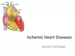
Myocardial Ischemia • Results when there is an imbalance between myocardial oxygen supply and demand • Most occurs because of atherosclerotic plaque with in one or more coronary arteries • Limits normal rise in coronary blood flow in response to increase in myocardial oxygen demand
Oxygen Carrying Capacity • The oxygen carrying capacity relates to the content of hemoglobin and systemic oxygenation • When atherosclerotic disease is present, the artery lumen is narrowed and vasoconstriction is impaired • Coronary blood flow cannot increase in the face of increased demands and ischemia may result
Ischaemic Heart Disease • Angina • Stable • Unstable • Prinzmetal’s • Myocardial Infarction • NSTEMI • STEMI
Angina • When ischemia results it is frequently accompanied by chest discomfort: Angina Pectoris • In the majority of patients with angina, development of myocardial ischemia results from a combination of fixed and vasospastic stenosis
Chronic Stable Angina • May develop sudden increase in frequency and duration of ischemic episodes occurring at lower workloads than previously or even at rest • Known as unstable angina: up to 70% patients sustain MI over the ensuing 3 months
Angina: cont • Patients with mild obstruction coronary lesions can also experience unstable angina • >90% of Acute MI result from an acute thrombus obstructing a coronary artery with resultant prolonged ischemia and tissue necrosis
Treatment of Angina • Treatment of Chronic Angina is directed at minimizing myocardial oxygen demand and increasing coronary flow • Where as in the acute syndromes of unstable angina or MI primary therapy is also directed against platelet aggregation and thrombosis
Epidemiology • Modifiable Factors: hyperlipidemia- ^ LDL (<130 normal) or low HDL (>60 normal), Hypertension, cigarette smoking and diabetes, obesity, BMI of >30 • Non-Modifiable Factors: advanced age, male sex, family medical history: male <55 y/o, female <65 y/o • Other: sedentary lifestyle and stressful emotional stress
Quality • Tightness, squeezing, heaviness, pressure, burning, indigestion or aching sensation • It is rarely “PAIN” • Never: sharp, stabbing, prickly, spasmodic, or pleuritic • Lasts a few seconds < 10 minutes • Relieved by NTG s/l • Levine Sign: clench fist to sternum
Signs & Symptoms accompany Angina • Dyspnea, nausea, diaphoresis resolve quickly after cessation of angina • Angina is a diffuse sensation rather than discrete
Ischemic Heart Disease • Imbalance between Myocardial oxygen supply and demand = Myocardial hypoxia and accumulation of waste metabolites due to atherosclerotic disease of coronary arteries
Stable Angina • Stable Angina: chronic pattern of transient angina pectoris precipitated by physical activity or emotional upset, relieved by rest with in few minutes • Temporary depression of ST segment with no permanent myocardial damage
Angina Pectoris • Angina Pectoris: uncomfortable sensation in the chest or neighboring anatomic structures produced by myocardial ischemia
Variant Angina • Typical anginal discomfort usually at rest • Develops due to coronary artery spasm rather than increase myocardial oxygen demand • Transient shifts of ST segment – ST elevation
Unstable Angina • Increased frequency and duration of Angina episodes, produced by less exertion or at rest = high frequency of myocardial infarction if not treated
Silent Ischemia • Asymptomatic episodes of myocardial ischemia • Detected by electrocardiogram and laboratory studies
Myocardial Infarction • Region of myocardial necrosis due to prolonged cessation of blood supply • Results from acute thrombus at side of coronary atherosclerotic stenosis • May be first clinical manifestation of ischemic heart disease or history of Angina Pectoris
Precipitants • Exertion: walking, climbing stairs, vigorous work using arms, sexual activity • Vasoconstriction: extremities, increased systemic vascular resistance, increased in myocardial wall tension and oxygen requirements • Myocardial Ischemia displays a circadian rhythm threshold for Angina it is lower in morning hours.
Physical Examination • Arcus senilis, xanthomas, funduscopic exam: AV nicking, exudates • Signs and symptoms: hyperthyroidism with increased myocardial oxygen demand, hypertension, palpitations • Auscultate carotid and peripheral arteries and abdomen: aortic aneurysm • Cardiac: S4 common in CAD, increased heart rate, increased blood pressure
Ischemia • Myocardial ischemia may result in papillary muscle regurgitation • Ischemic induced left ventricular wall motion abnormalities may be detected as an abnormal precordial bulge on chest palpation • A transient S3 gallop and pulmonary rales = ischemic induced left ventricular dysfunction
Diagnostic Tests • Blood tests include serum lipids, fasting blood sugar, Hematocrit, thyroid (anemias and hyperthyroidism can exacerbate myocardial ischemia • Resting Electrocardiogram: CAD patients have normal baseline ECGs • pathologic Q waves = previous infarction • minor ST and T waves abnormalities not specific for CAD
Electrocardiogram • Electrocardiogram: is useful in diagnosis during cc: chest pain • When ischemia results in transient horizontal or downsloping ST segments or T wave inversions which normalize after pain resolution • ST elevation suggest severe transmural ischemia or coronary artery spasm which is less often
Exercise Stress Test • Used to confirm diagnosis of angina • Terminate if hypotension, high grade ventricular disrrhythmias, 3 mm ST segment depression develop • (+): reproduction of chest pain, ST depression • Severe: chest pain, ST changes in 1st 3 minutes, >3 mm ST depression, persistent > 5 minutes after exercise stopped • Low systolic BP, multifocal ventricular ectopy or V- tach, ST changes, poor duration of exercise (<2 minutes) due to cardiopulmonary limitations
Other Diagnostic Tests • Radionuclide studies • Myocardial perfusion scintigraphy • Exercise radionuclide ventriculography • Echocardiography • Ambulatory ECG monitoring • Coronary arteriography
Management Goals to reduce Anginal Symptoms • Prevent complications – myocardial infarction, and to prolong life • No smoking, lower weight, control hypertension and diabetes • Patients with CAD – LDL cholesterol should achieve lower levels (<100) • HMG-COA reductase inhibitors are effective
Pharmacologic Therapy • Therapy is aimed in restoring balance between myocardial oxygen supply and demand • Useful Agents: nitrates, beta-blockers and calcium channel blockers
Nitrates • Reduce myocardial oxygen demand • Relax vascular smooth muscle • Reduces venous return to heart • Arteriolar dilators decrease resistance against- which left ventricle contracts and reduces wall tension and oxygen demand
Nitrates: cont • Dilate coronary arteries with augmentation of coronary blood flow • Side effects: generalized warmth, transient throbbing headache, or lightheadedness, hypotension • ER if no relief after X2 nitro's: unstable angina or MI
Problems with Nitrates • Drug tolerance • Continued administration of drug will decrease effectiveness • Prevented by allowing 8 – 10 hours nitrate free interval each day. • Elderly/inactive patients: long acting nitrates for chronic antianginal therapy is recommended • Physical active patients: additional drugs are required
Beta Blockers • Prevent effort induced angina • Decrease mortality after myocardial infarction • Reduce Myocardial oxygen demand by slowing heart rate, force of ventricular contraction and decrease blood pressure
Beta -1 • Block myocardial receptors with less effect on bronchial and vascular smooth muscle- patients with asthma, intermittent claudication
Beta-Agonist blockers • With partial B-agonist activity: • Intrinsic sympathomimetic activity (ISA) have mild direct stimulation of the beta receptor while blocking receptor against circulating catecholamines • Agents with ISA are less desirable in patients with angina because higher heart rates during their use may exacerbate angina • not reduce mortality after AMI
Beta-blockers • Duration of beta-blockers depends on lipid solubility • Accounts for different dosage schedules
Contraindications • Symptomatic CHF, history of bronchospasm, bradycardia or AV block, peripheral vascular disease with s/s of claudication
Side Effects • Bronchospasm (RAD), CHF, depression, sexual dysfunction, AV block, exacerbation of claudication, potential masking of hypoglycemia in IDDM patients
Calcium Channel Blockers • Anti-anginal agents prevent angina • Helpful: episodes of coronary vasospasm • Decreases myocardial oxygen requirements and increase myocardial oxygen supply • Potent arterial vasodilators: decrease systemic vascular resistance, blood pressure, left ventricular wall stress with decrease myocardial oxygen consumption
Calcium Channel Blockers • Secondary agents in management of stable angina • Are prescribed only after beta blockers and nitrate therapy has been considered • Potential to adversely decrease left ventricular contractility • Used cautiously in patients with left ventricular dysfunction
Drug Selection • Chronic Stable Angina: beta blocker and long acting nitrate or calcium channel blocker (not verapamil: bradycardia) or both. • If contraindication to BB a CCB is recommended (bronchospasm, IDDM, or claudication) any of CCB approved for angina are appropriate.
Drugs • Verapamil and Cardizem is preferred because of effect on slowing heart rate • Patients with resting bradycardia or AV block, a dihydropyridine calcium blocker is better choice • Patients with CHF: nitrates preferred amlodipine should be added if additional therapy is needed
Drugs • Primary coronary vasospasm: no treatment with beta blockers, it could increase coronary constriction • Nitrates and CCB are preferred • Concomitant hypertension: BB or CCB are useful in treatment • Ischemic Heart Disease & Atrial Fibrillation: treatment with BB, verapamil or Cardizem can slow ventricular rate
Combination Therapy • If patients do not respond to initial antianginal therapy – a drug dosage increase is recommended unless side effects occur. • Combination therapy: successful use of lower dosages of each agent while minimizing individual drug side effects