Download
1 / 17
170 likes | 307 Views
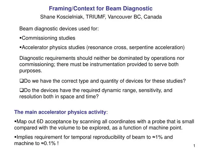
Framing/Context for Beam Diagnostic. Shane Koscielniak, TRIUMF, Vancouver BC, Canada. Beam diagnostic devices used for: Commissioning studies Accelerator physics studies (resonance cross, serpentine acceleration).

E N D
Framing/Context for Beam Diagnostic Shane Koscielniak, TRIUMF, Vancouver BC, Canada • Beam diagnostic devices used for: • Commissioning studies • Accelerator physics studies (resonance cross, serpentine acceleration) Diagnostic requirements should neither be dominated by operations nor commissioning; there must be instrumentation provided to serve both purposes. • Do we have the correct type and quantity of devices for these studies? • Do the devices have the required dynamic range, sensitivity, and resolution both in space and time? • The main accelerator physics activity: • Map out 6D acceptance by scanning all coordinates with a probe that is small compared with the volume to be explored, as a function of machine point. • Implies requirement for temporal reproducibility of beam to ≈1% and machine to ≈0.1% !
The core functions are to measure/reconstruct as follows • Thread along beamlines • First turn – includes inject/extract • Measure beam transmission (always, always, always). • Find closed orbits and optics functions (α,β,φ,δ) versus momenta. • Find ToF or revolution frequency versus momenta. • Map out acceptance (6D) • Verify probe emittance (6D) Beam Transmission (resolution 1% or better) 2 Fast Faraday cups in injection/ERLP and extraction lines are essential for calibrating other non-intercepting devices. However they are beam destructive. Resistive wall-current monitor in ring (turn by turn) BUT for how many turns? RWCM or current toroids in injection/extraction lines? Faraday cups can be calibrated to 0.1% with precision current measurer.
Thread along inject & extract beamlines H&V BPM or screen/wire near all magnetic elements (exclusive OR) • First turn. Needs: • 1 screen immediately before and after injection point (i.e. 2 screens total) • 1 screen immediately before and after extraction point (i.e. 2 screens total) • ≈ 42 H&V BPMs in ring, with single passage acquisition. • Have to synchronize kicker(s) firing with beam arrival. • Therefore timing pulse either from ERLP RF and/or beam in injection line from WCM. Play with ΔT offset and kicker strength until you see spot on the screen.
Function: Closed orbits and optics functions versus momentum • Devices: BPMs (local accuracy ±25 μm equivto 50μm resolution) • BPMs are the most crucial piece of instrumentation. • Measure static closed orbits as function of injection energy and reconstruct optics functions including phase advance per cell as sampled at discrete locations. 5-turn algorithm to start and 1024 turn memory for later. • Finding the golden orbits (i.e. finding closed orbit correction across a range of momenta) is a major commissioning activity. • An algorithm for golden orbits has been proposed. • Can the optimum accelerated orbit be constructed from static measurements? It may be the only way! • Needs: • H&V BPMs at every opportunity • Single-turn time resolution, and many-turn acquisition/storage for each BPM. • Dynamic range: not an issue, will always work with 1-2 E8 electrons. • Resolution: ≈ 50 μm across 2-3 cm
Transverse Acceptance – use BPMs One can think to measure the acceptance at some particular momentum, and the acceptance of an accelerated orbit. Scan the probe emittance in (x,x’, y,y’) and reconstruct betatron oscillations in the ring and extraction line. If the optics functions are known (from commissioning), then you can reconstruct the swept out emittance – up to the MC acceptance. Horizontal scanning by external dipole and kickers within the ring. Vertical scanning by 2 external dipoles. Vertical COD correction of drifts by 16(?) circuit-board type dipoles? Verification of probe emittance at injection and extraction – use transverse profile monitors (resolution 50-100μm, depends on β function) This is reconstructed from 3 H&V measurements of beam profile, either with wires or harps or screens. In principle, the ideal is two sets of identical measurement rigs in the injection and extraction lines – with similar phase advances. See 26 Feb 2007 talk for pros & cons of wires versus harps versus screens, etc.
Probe transverse emittance - continued However, the measurement is destructive. Hence one cannot measure “in one shot” the “before and after” probe emittance. An outrageous cost-saving measure? Shall we put it to rest? Hence, if one can assume that passage through a partial machine turn is not damaging, could measure both the injected and extracted probe emittance in the extraction line. May have to reset the beamline optics between pulses to do this. BUT have to thread beam through machine first! There is also opportunity, for small number of turns, to see change of probe emittance via OTR screens or wires in the ring – gives profiles and emittance is inferred. These same screens (2) are essential for commissioning. Requirement for turn-by-turn operation: low spot remanence, fast shutter or large turn separation. Screen driven in from outside of ring. Issue: resolution. Inside the ring, smallest beam width is ≈0.3mm. (β function 0.2 to 0.6 m) Resolving sub-structure will be difficult for moving wire or OTR. This makes questionable their use in the ring except for commissioning. Pitch/Step size 50 μm, wire size 10μm.
Longitudinal Acceptance Assumption: small probe is used to map out acceptance. ERLP has quoted a variety of bunch length and momentum spread. We shall work with an emittance of ≈ 1 π keV.ns. (i.e. FW 100 keV, 40 ps), but Muratori has quoted smaller values down to FW 20 keV, 15 ps – which is certainly a small probe! Acceptance mapping Sweep RF-phase and energy of the injected beam; and measure these same quantities. Implies desired variability of ERLP extraction energy – AND sophisticated RF synchronization system Then kick out after 1,2,3…. Turns to extraction line RF-phase and frequency Exploration of the acceptance and of “different machines”, implies the ability to synchronize the phase (with a given, variable offset) of the EMMA ring to the phase of the ERLP beam (for which one may substitute EMMA RF), while the EMMA ring operates across a narrow band of RF.
1st coordinate is beam phase measured in ring and extraction line • Centroid phase (or Δt) can probably be measured to 2o or better. • What device? • w/b resistive wall-current monitors (1 in ring, 1 in extraction line) • or Σ signal from BPMs. These (capacitive buttons) give differentiated signal. Is zero crossing or other property good measure of RF phase? 2nd coordinate of probe centroid is its momentum Ideally: identical spectrometer magnet (Hall probe), slits and Farday cup in injection and extraction lines. (resolution 0.1%) In addition to measuring probe input/output values, this system could be used to calibrate the closed orbits versus momentum during commissioning. The momentum measurement is beam destructive. Hence cannot simultaneously find inject/extract momenta. Hence, if one can assume that passage through a partial machine turn is not damaging (i.e. RF off), could measure both the injected and extracted probe energy in the extraction line. Or use the dipole and Faraday cup at end of ERLP first straight. (Assumes cup is moveable or dipole has independent PS. Is this true?
Verification of probe longitudinal emittance • Momentum spread of probe measured as above with high resolution spectrometer. • Longitudinal profile of probe (resolution 4o or better at 1.3 GHz) • Short bunch (<40 ps), therefore need very large band-width. • Options (in extraction line): • Transverse deflecting mode cavity. • Opto-electronic (Faraday rotation, etc). • Ratio of two Fourier components. Model depenent, but workable. Need two RF cavities, at 1.3 GHz and, say, 2.6 GHz. Gives less information than 1) or 3) but maybe much less costly. Passive cavities are used as beam pick-ups. • In all cases, need initial beam on axis. Hence this measurement incompatible with simultaneous transverse acceptance study. • May need steering magnets in extraction line, or quadrupole on movers?! NB, we refer here not to resolving the centroid phase, but to resolving changes in the bunch length, or even substructure within the bunch.
Time of Flight (ToF) versus momentum With the RF off, the ToF variation is up to 3-4 ps/cell. This amounts to a wopping 120-160 ps per turn. (56 to 75 deg of RF phase at 1.3 GHz). This kind of phase slip against a standard 1.3 GHz clock, should be easy to measure in the time domain in a single-turn measurement. Suspect the the resolution is better than 1 ps, so you could measure in 1% steps of momentum quite comfortably. If it were not for beam loading and debunching, could arbitrarily increase precision by waiting more turns. Another possibility is to measure with RF on, and in frequency domain. This has advantage that we want to measure transverse oscillations for up to 1000 turns, and that requires a bunched beam. For beam in bucket, ΔT=nτ+δTCos(Ωt) and the smaller is the synchronization error δT, the closer is the ToF per turn to harmonic#/RF. Trouble is we wanted the ToF of the beam for a particular energy, not to measure the known RF period! What is needed is to make a conventional frequency lock of the RF to the beam without changing (much) the bunch energy or arrival time. After lock-in, RF has become equal to h/ToF; and RF is easily measured with a frequency meter. Can we make this fast enough for it to be useful? What to use for phase probe? BPM or WCM?
Wires or harps or screens? Scanning wire = single wire, resolution depends on step size Harp = set of wires strung on a frame, resolution depends on pitch size Screen = 2D flat surface, resolution depends on optics/magnification and size off CCD camera array. Wires and harps measure electrons arriving or leaving, or at HEP gammas via scintillator. Need 2 devices to get H&V coverage. Screens are luminescent, so you count photons. 2D image directly. Wire takes many beam repetitions to build picture. Harps and screen get whole picture in one shot. In beamlines, issues of remanence and shuttering are irrelevant. Wire has, in principle, enormous dynamic range, 103 or more. Screens come as YAG or OTR. YAG saturates and is non-linear, whereas OTR is a proportional device that can give beam distribution. Do OTRs work well at 10 MeV and with charge down to 1E8? Issue is whether there is a lower limit of sensitivity, to charge/pixel of the screen. If not, I would prefer the OTR.
Waveguide distribution IOT x 3 (2) Septum 72° Kicker Kicker Kicker power supplies Septum 72° Wire Scanner Cavity x 19 Kicker Screen Kicker EMMA Screen Kicker power supplies D Quadrupole x 42 F Quadrupole x 42 BPM x 82 Wire Scanner Locations for 16 Vertical Correctors Wall Current Monitor
INJECTION LINE In ERLP New Dipoles x 2 (32.5°) & BPMs at dipole entrance Spectrometer Screen & Faraday cup Screen x 3 - Tomography Section Screens or Wire Scanners ? Vertical steering to be added ! Screen & BPM Wall Current Monitor New Quadrupoles x 11 Screen & Vert. Slit New Dipole 30° & BPM at dipole entrance Vacuum valve Screen Bunch length measurement? Kicker Kicker SRS Quadrupoles x 6 Septum 72° ERLP Faraday Cup Vacuum valve EMMA New Dipole 30° OTR Screen in ERLP Quadrupoles x 2 in ERLP ERLP
Screen & BPM SRS Quadrupoles x 3 Screen & Vert. Slit Screens or Wire Scanners ? Spectrometer (extracted momentum) Screen BPM @ dipole entrance F/Cup Vacuum valve New Dipole 21° & BPM at dipole entrance Screen New Quadrupole x 10 Transverse Deflecting Cavity (longitudinal profile) or E-O Monitor Screen x 3 Tomography Section (emittance measurement) Wall Current Monitor
Discussion topics • Does commissioning needs add extra beam diagnostic or other devices? • What are the “extra” screens and vertical slits for? In inject/extract lines. Have no real objection, but maybe they are replacable by BPMs? • Why does it say bunch length measurement on inject line? • Is it acceptable to perform most of the beam diagnostic in the extraction line, and eliminate some devices from the injection line as a cost saving measure? Or are these too far apart for comfort? Beam properties measured in ½ pass through ERLP were considered “too far away” on Monday. Maybe think of this as a cross-calibration exercise. • Proceed with opto-electronic, or investigate passive cavities? • Should we add a full complement of BPMs to beamlines? • Wires or OTRs in beamlines for tomo? • What to use as phase probe? BPM or WCM? What is the optimum phase advance between profile monitors for transversal emittance reconstruction? For example, tomographic argument would say 60o. Should the beta-functions be larger for better resolution?
Aberrations Not mentioned yesterday, but its something we shall measure. ToF is not a line, it has thickness depending on betatron amplitude. Chromaticity = Plot of tune versus momentum. Betatron tune versus betatron amplitude. Process BPM data for long enough? ToF versus betatron amplitude, how do we construct this? What series of measurements is needed?