Download

1 / 46
490 likes | 734 Views
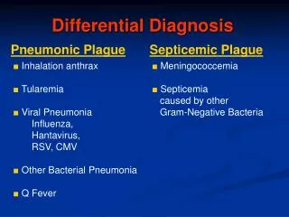
Differential Diagnosis. Staphylococcus aureus. Mycobacterium marinum. Vibrio vulnificus. Ecthyma gangrensum. Aeromonas. Deep fungal infection. Anthrax. Erysipeloid. Orf. Tularemia. vasculitis. Diagnostic Procedure(s) and Result(s).

E N D
Diagnostic Procedure(s) and Result(s) • Cultures of the lesion on the left leg and right hand, obtained on admission, grew rare methicillin-susceptible Staphylococcus aureus colonies; the culture of the leg also grew Enterococcus in the thioglycollate broth tube, which was thought to be a contaminant.
Cultures of the leg and right hand obtained in the operating room were sterile.
All other wound cultures, including for fungi and mycobacteria, were sterile, as were blood cultures. Testing for 1, 3 beta D glucan was negative.
Indirect immunofluorescence testing for anti-neutrophil cytoplasm antibodies (ANCA), serum protein electrophoresis and testing for lupus anticoagulant and syphilis were also negative.
Histopathological examination • revealed normal tissue, as well as an intraepidermal blister, a dense dermal acute inflammatory infiltrate and tissue necrosis.
Pyoderma gangrenosum is an idiopathic inflammatory disease that is often mistaken for an infectious process.
Diagnosis of this condition is based upon clinical history, pathology, and exclusion of other diseases that cause erosive or ulcerative skin lesions.
While an underlying systemic disease such as inflammatory bowel disease, myeloproliferative disorder, or inflammatory arthritis is found in approximately 50% of cases of pyoderma gangrenosum, this disease has also been associated with trauma (including burns) and surgery.
It is important to consider this disease in patients with non-healing wounds and obtain early dermatologic consultation whenever possible, because surgical intervention, including debridement, may exacerbate pyoderma gangrenosum and lead to worsening ulceration.
While there is no published algorithm for the treatment of pyoderma gangrenosum, the literature suggests the use of prolonged systemic glucocorticoid therapy;
A subset of patients requires the use of other immunomodulators (e.g. cyclosporine, thalidomide, and tumor necrosis factor (TNF)-alpha blockers such as infliximab).