Download
1 / 19
210 likes | 323 Views
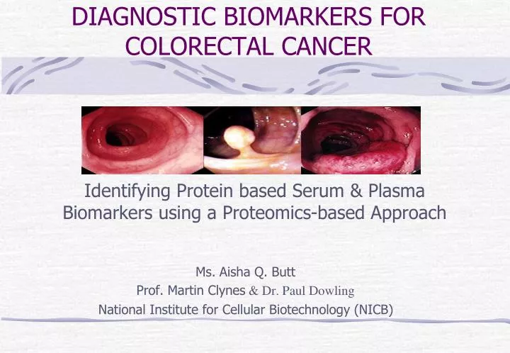
DIAGNOSTIC BIOMARKERS FOR COLORECTAL CANCER . Identifying Protein based Serum & Plasma Biomarkers using a Proteomics-based Approach. Ms. Aisha Q. Butt Prof. Martin Clynes & Dr. Paul Dowling National Institute for Cellular Biotechnology (NICB). Objectives.

E N D
DIAGNOSTIC BIOMARKERS FOR COLORECTAL CANCER Identifying Protein based Serum & Plasma Biomarkers using a Proteomics-based Approach Ms.Aisha Q. Butt Prof. Martin Clynes & Dr. Paul Dowling National Institute for Cellular Biotechnology (NICB)
Objectives • Colorectal Cancer: Facts & Diagnostic Limitation • Hypothesis & Aims • Research Strategy • Biomarker Discovery • Cell Culture • Nano-HPLC & Mass Spectrometry • Bioinformatics • Biomarker Verification • ELISA • Immunohistochemistry • Conclusion & Future Work
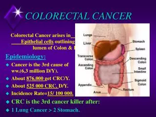
Colorectal Cancer: Facts & Figures • 1 Million new cases & 1/2 million deaths p/yr • 3rd most common malignancy in world. • Primary Cancer mortality cause in: USA, Europe & Korea • 2nd most common cause of death in Republic of Ireland. • Approximately, 2000 new CRC cases diagnosed each year. • Ireland: Highest bowel cancer incidence, lower survival rate and higher mortality rates in Western Europe both in men and women. • Prediction: By 2020, new CRC cases projected to ↑ by 79% in men & 56% in women.
CRC Diagnosing Limitations Our Hypothesis & Aims Devise screening programme based on Panel of Protein Biomarkers Used in combination with Colonoscopy To offer most accurate test for early diagnosis of CRC Novel high value biomarker diagnostics test kit based on blood or serum could: Prescreen high risk groups Eliminate waiting lists Permit rapid access to suspects • CRC highest cure rates if detected early. • Colonoscopy • Gold standard for diagnosis of colonic neoplasia • But it’s not a perfect test • Lengthy waiting lists for colonoscopy services in Ireland • Lack of sensitivity & low compliance – Most of current diagnostics detect cancer after it has already spread to other parts of the body
Research Strategy • Cancer Secretome: comprising all proteins released by tumour cell – Attracting much attention recently. • Our Approach – Collection of Conditioned Media from 4 CRC cell lines HCT116, HT-29, SW480 & WiDr using Cell Culturing Techniques. • Proteins identified from conditioned media using Mass Spectrometry– evaluated using specific criteria to identify most likely candidate for further investigations. • Criteria: Being detectable in all cell lines • Likelihood that proteins are shed/secreted by tumour cells • Proteins primary localisation & molecular function within the cell • Literature search • Bioinformatics allowed data sets to be cross compared & from the shortlisted data, few proteins were verified using: • ELISA kit • Immunohistochemistry
Biomarker Discovery • Cell Culture • Four CRC cell lines cultured in respective media- HCT116, HT-29, SW480 & WiDr • Assessment of Cell Viability performed on derived Conditioned Media • Protein Concentration • Proteins in CM’s centrifuged down in CENTRICONS (Sartorius Stedim Biotech) • Concentrated proteins by 40 fold (4mls to 100µl) • Protein Precipitation & Clean Up • Using Ready PrepTM 2-D Clean-Up Kit protein samples subjected to precipitation and cleaning using buffers and reagents yielding ~30µl sample for each cell line • In Solution Digestion / Double Digestion • Double Digestion carried out using LysC & Trypsin for denaturation & digestion of proteins into peptides. • Sample Clean Up • Using PepClean TM C-18 Resin Spin Columns, tryptic peptide mixtures were further cleaned up for removal of any solvents and buffers. • Sample speed vacuumed down and resuspended in Mass Spec. compatible buffer.
LC-MS & Bioinformatics • Ettan MDLC system (GE Healthcare, Piscataway, NJ) - applied for desalting and separation of LysC and tryptic peptide mixtures. • Two experiments set up: • 10µl tryptic peptide sample loaded onto LC column with 1hour gradient time • 20µl tryptic peptide sample loaded onto LC column with 3hour gradient time • MDLC-MS results analysed using Bioworks Browser software suite TM (Thermo Fisher Scientific, USA) • Proteins in samples searched against Swiss Prot human protein database (Dec, 2009; 65,533 entries; 20,339 human proteins). • The stringent protein identification criteria based on multiple filters: • Distinct peptides • Delta CN (0.040) • Xcorr vs. Charge state (1.50, 2.20, 2.50, and 3.00) • Peptide Probability (0.05) • Number of distinct peptides (2)
Results & Discussion • Conditioned Media – Cell Viability • Mean Average: 89.875% Viable Cells & 10.125% Dead Cells • Cell counts performed using haemocytometer and trypan blue dye on CM derived from 4 CRC cell lines using NICB-SOP-003-01.
Mass Spectrometry – Sample Gradient Time • An approximate >2.5 fold increase in the total proteins identified on MS using 1hr vs. 3hr sample gradient time. • Stringent Multiple filters applied: Distinct Peptides, Delta CN (0.040), Xcorr vs. Charge state (1.50, 2.20, 2.50, and 3.00), Peptide Probability (0.05) and Number of distinct peptides (2). • Data Cross compared using Bioinformatics & Excel tools: • 92 common proteins identified within 4 CRC cell lines.
Cellular Localisation & Biological Process • 92 Common proteins further analysed & classified using Human Protein Reference Database on basis of: • Cellular Localisation • Biological Processes • Motifs • 51% Proteins localization is based within cytoplasm and 23% proteins are associated with cell growth & maintenance. • 92 Common proteins further analysed & classified using Human Protein Reference Database on basis of: • Cellular Localisation • Biological Processes • Motifs • 51% Proteins localization is based within cytoplasm and 23% proteins are associated with cell growth & maintenance.
Protein Motifs • Proteins secreted/shed by the tumour cells (and so will be detectable in the circulatory system) – an important criterion to identify the most likely candidate biomarkers for further investigations. • Proteins having signal peptide on their motifs are secretory proteins, whereas all other proteins are shed by the tumour cells.
Biomarker Verification • Stringent classification of 92 common proteins on the basis of: • Protein’s Cellular Localisation • Biological Process • Protein’s Motifs • Allowed to choose 2 proteins: • Protein X • Protein Y • Why these 2 Proteins? • Presence of signal peptide motif on Protein X • Relevance and association of Protein Y with other cancer types (Literature Search)
ELISA Test – Serum & Plasma Samples • Protein X levels in serum & plasma samples measured using double-antibody sandwich ELISA system (Bender MedSystems, Austria) • Serum samples: 16 Advanced stage + 8 Healthy controls • Plasma samples: 9 Early stage + 8 Healthy controls (Early Stage )
Receiver Operating Characteristics – ROC Curve • ROC curve analysis of Protein X for discriminating CRC patients from healthy controls for advanced stage serum (AUC - 0.825) and early stage plasma samples (AUC - 0.639). Protein X Diagnostic efficacy in Serum samples Protein X Diagnostic efficacy in Plasma samples
Immunohistochemistry – CRC Tissue Sections • CRC tissue sections compared to normal colon tissues via Protein Y expression using Immunohistochemical staining. • Protein Y positive expression observed in a moderately differentiated adenocarcinoma (intermediate differential grade) • Protein Y positive staining in the cytoplasm - Granular in nature • A lot less nuclear staining in areas of tumour compared to normal tissue
Conclusion • This pilot study has shown that cancer secretome from the tumour cells presents a promising reservoir of biomarkers with soluble-secreted proteins and shed membrane proteins. • And, the use of these secreted proteins to go back on clinical serum or plasma samples to distinguish patients with or without CRC is a promising diagnostic approach. • Also, both Protein X and Protein Y could serve as potential biomarkers if used in combination with few other biomarkers for the early diagnosis of colorectal cancer.
Further Work... • Further work and efforts are required to fully validate the biomarkers detected in this study with: • Large sized clinical samples • Multiple medical centre samples • ELISA Test for Protein Y protein on serum & plasma samples • These biomarkers detected can be used in combination with a panel of other protein biomarkers and colonoscopy to improve the overall accuracy and speed for detecting CRC at an early stage and prioritise high-risk individuals.
Summary • Colorectal Cancer & Diagnostic Limitations • Aim: Protein based Biomarkers for early detection of CRC • Biomarker Discovery: Research Strategy • Cell Culture • Nano-HPLC & Mass Spectrometry • Bioinformatics • Biomarker Verification • ELISA • Immunohistochemistry • Conclusion & Future Work