Download
1 / 41
430 likes | 715 Views
Ch. 6: Bone & Skeletal Tissue Skeletal Cartilages Classification of Bones Functions of Bones Bone Structure Development Bone Homeostasis: Remodeling & Repair Homeostatic Imbalances of Bone Developmental Aspects. Ch. 6: Bone & Skeletal Tissue Skeletal system includes bone & cartilage

E N D
Ch. 6: Bone & Skeletal Tissue Skeletal Cartilages Classification of Bones Functions of Bones Bone Structure Development Bone Homeostasis: Remodeling & Repair Homeostatic Imbalances of Bone Developmental Aspects
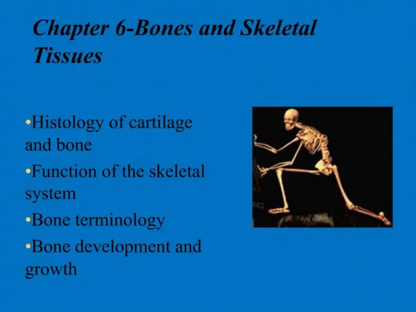
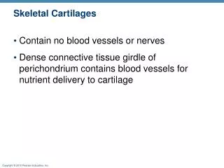
Ch. 6: Bone & Skeletal Tissue Skeletal system includes bone & cartilage Skeletal Cartilages Structure, types, locations All types of sk cart have chondros, extracell mtx, fibers Hyaline c most common, flexible, resilient, contain fine collagen fibers Articular, costal, respiratory (vb), nasal (ext nose) Elastic c like hyaline; more elastic fibers; tolerate repeated bending; epigolttis, ext ear Fibrocart Cross between hyal cart (chondros) & dense reg conn tiss (thick coll fibers); high tensile strength Resist high pressure & stretch Menisci in knees, intervert discs Growth Appositional; Interstitial
Forces • Tension • Compression • Bending Ch. 6: Bone & Skeletal Tissue Classification of Bones Axial vs appendicular By shape: long, short, flat, irregular Functions of Bones Support Protection Movement Storage of minerals, growth factors, fat Hematopoesis
Ch. 6: Bone & Skeletal Tissue Bone Structure Gross Anatomy Bone Markings – See Table 6.1 Texture Compact (lamellar): dense outer layer, naked eye smooth Spongy (cancellous): trabeculae, red or yellow marrow Typical long bone structure Diaphysis: compact bone collar over marrow-filled medullary cavity Epiphyses: bone ends, compact over spongy, articular (hyaline) cartilage; epiphyseal line (remnant of epiphyseal plate) Membranes: periosteum, endosteum, osteoblasts/clasts Short, irregular, flat bone structure Compact over spongy; no marrow cavity Hematopoetic tissue (red marrow): Trabecular parts of long bones (esp. heads of femur, humerus in adult) & flat bones Microscopic Anatomy Chemical Composition
Ch. 6: Bone & Skeletal Tissue Bone Structure Gross Anatomy Microscopic Anatomy Cell types: osteogenic, osteoblasts, osteocytes, osteoclasts Compact bone Microscopic passageways; osteons (Haversian systems) Multiple lamellae per osteon; alternating collagen fiber orient’n Central canal; osteocytesbtn lamellae Spongy bone Trabeculae align with stress; no osteons Chemical Composition (skeleton = 14-17% of total adult body weight i.e. ~ 10 kg) Organic Cells & osteoid (ground substance & collagen fibers) Inorganic(2/3 of skeleton weight – Trotter & Peterson, J Bone JtSurg Am 1962) Hydroxyapatite crystals (mainly calcium phosphate)(40% of hydroxyapatite is Ca; total body Ca ~ 2.5 – 3 kg for normal 70 kg person)
Bone architecture (a) macroscopic bone, (b) osteons with circular arrangements of differently oriented collagen fibers. (c) collagen fiber = bundles of collagen fibrils. (d) collagen fibril = staggered arrangement of collagen molecules with embedded mineral crystals (blue). (e) collagen molecule triple helix. Collagen: tensile and bending strength. Hydroxyapatite (calcium phosphate): compressive strength Reinforced concrete “Rebar” (steel reinforcing bars): tensile and bending strength. Concrete:compressive strength. Above: channel.nationalgeographic.com/series/la-hard-hats/all/Photos#tab-Photos/11 Left: www.bnl.gov/nsls2/sciOps/chemSci/softMatter.asp College or Department name here
Ch. 6: Bone & Skeletal Tissue Development Ossification (= osteogenesis) Forming the bony skeleton Intramembranous ossif Endochondral ossif Postnatal bone growth Length of long bones: epiphyseal plate Hormonal regulation GH. Sex hormones: growth spurt, epi plate closure
Ch. 6: Bone & Skeletal Tissue Bone Homeostasis: Remodeling & Repair Remodeling At periosteum & endosteum Remodeling unit w/ osteoblasts, osteoclasts Osteocytes (stimulated by osteoblasts) secrete matrix Control of remodeling By hormones to regulate plasma [Ca] PTH ↑when [Ca] ↓, stimulates osteoclasts In response to mechanical stress Bone strongest where stress acts Long bone hollow ctr; shaft thickest at middle Curved bones thickest where most likely to break Spongy bone trabeculae line up along stress lines Projections (tuberosity, crest,…) where muscles attach
Ch. 6: Bone & Skeletal Tissue Bone Homeostasis: Remodeling & Repair Remodeling animation with description of osteoporosis (41 s) http://www.youtube.com/watch?v=NKVKNqIOnh0&feature=related Another, similar (54 s): http://www.youtube.com/watch?v=5uAXX5GvGrI
Ch. 6: Bone & Skeletal Tissue Bone Homeostasis: Remodeling & Repair Repair Classification of fractures Bone end positions: displaced or nondisplaced Complete or incomplete Orientation: linear (long axis) or transverse Skin penetration: open (compund) or closed (simple) By nature, location of break – see Table 6.2 Repair process – see Fig 6.15
A current debate: Do some osteoporosis drugs increase the rate of “rare” fractures? Radiograph Showing a Subtrochanteric Stress Fracture. Kwek et al., NEJM 359:316, 2008. Not the most common site for stress fracture. Common stress fracture sites: tibia (runners), metatarsals (dancers, skaters), fibula…) From article about possible increase in fracture risk in patients on bisphosphonates (osteoporosis drugs). See also JAMA 305: 783-789, 2011. Some data suggests that the risk of rare fractures is a lot higher after 6-8 years on bisphosphonates than after 2 yrs; some suggest a bisphosphonate holiday after 5 yrs (NYT 2011-03-06).
Ch. 6: Bone & Skeletal Tissue Homeostatic Imbalances of Bone Osteomalachia Failure to mineralize; weak bones Rickets in children Due to Ca deficiency, maybe secondary to vit D deficiency Osteoporosis Resorption > formation; ↓ bone density, mass Elderly susceptible; vertebral, femoral neck fx Estrogen, testosterone slow osteoclasts, stimulate blasts Post-menopausal fall in estrogen Remodeling animation with description of osteoporosis (41 s): http://www.youtube.com/watch?v=NKVKNqIOnh0&feature=related Paget’s disease Disorganized (Pagetic) bone; spongy/compact too high Elderly susceptible; affected bone weak
Hip Fractures • 300,000 hip fractures annually in US • Majority related to osteoporosis and falls in olderpeople • Enormous public health implications andeconomic burden • A topcause of immobilization in elderly • Patients who have sustained a hip fracture • 2-year mortalityrate of 36% • Immobility → increasedbone resorption, predisposition to 2nd hip fracture • Many don’t regain prefracture level of mobility; lose independence, QOL • Risk of subsequent skeletal fracture up 2.5x • Risk of new hipfracture up 5x – 10x Editorial: Calis KA, Pucino F, NEJM 2007: Sep 17, epub ahead of print
Further reading: NYTimes Sep 23 2009 Can vitamin D improve athletic performance? http://well.blogs.nytimes.com/2009/09/23/phys-ed-can-vitamin-d-improve-your-athletic-performance/ Vitamin D essential for absorption of Ca from gut; it also helps other cells including muscle cells use Ca. Many of us with a “normal” diet and occasional outdoor time are vitamin D deficient and we may benefit from more D. Vitamin D Status and Its Relation to Muscle Mass and Muscle Fat in Young Women.59% of sample (16-22 yo ♀ in Calif) had insufficient 25OH-D (<30 ng/ml), including 24% who were deficient in 25OH-D (<20 ng/ml) (surprisingly large %). Vitamin D insufficiency was associated with increased fat infiltration in muscle. Gilsanz, …, Kremer. J Clin Endocrinol Metab 2010; DOI: 10.1210/jc.2009-2309
Further reading: NY Times Sep 1 2009 Does ibuprofen help or hurt during exercise? http://well.blogs.nytimes.com/2009/09/01/phys-ed-does-ibuprofen-help-or-hurt-during-exercise/ Many athletes regularly take ibuprofen. Article describes increasing evidence that "vitamin I" has negative side effects including blunted growth response of bone to applied stress, decreased post-workout collagen production, etc. One scientist wrote recently in Brit J Sports Med that regular ibuprofen use around the time of exercise diminishes the expected increase in skeletal strength that otherwise comes from exercise. http://bjsm.bmj.com/cgi/content/full/43/8/548