Download
1 / 27
270 likes | 280 Views
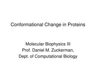
Conformational Change in Proteins. Molecular Biophysics III Prof. Daniel M. Zuckerman, Dept. of Computational Biology. Conformational Change & Function. Many (most?) proteins function via conformational changes Outline Ensemble Picture and Examples Hemogolobin and allostery

E N D
Conformational Change in Proteins Molecular Biophysics III Prof. Daniel M. Zuckerman, Dept. of Computational Biology
Conformational Change & Function • Many (most?) proteins function via conformational changes • Outline • Ensemble Picture and Examples • Hemogolobin and allostery • Myosin, kinesin and motion • Functional motif for NTP hydrolysis • Calmodulin and signalling • Reference/reading: Berg et al., Biochemistry • Also source for figures
intermediates structural ensemble B structural ensemble A Ensemble Picture (Statistical Mechanics) • An ensemble of paths, traversing ensembles of intermediate structures, connects two ensembles of structures
Motor proteins Myosin
Fluctuations in Biology • Regulation: fatty acid binding proteins
Enzyme Conformational Change • Adenylate kinase Open/ligand-free Closed: Ligand-bound
Calcium-bound Calcium-free Calmodulin, N-terminal lobe Conformational Change & Signaling • Signaling protein: Calmodulin
More dramatic conformational flexibility • Open and closed Ca-bound calmodulin • Likely both occur in solution … and everything “in between” Calcium-bound Calcium-bound
Consequences of Induced Fit Idea • The idea: Ligand binding induces conformational change • Some possibilities: • Ligand binds to an apo-like or holo-like configuration • Ligand unbinds from holo-like or apo-like configuration • One way or another, proteins must undergo large conformational fluctuations • And this must happen all the time to allow constant binding and un-binding
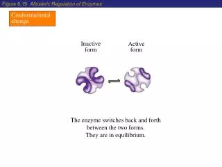
Allostery: “cooperativity” in binding • For proteins with more than one binding site, the binding events often are not independent • Even when binding sites are identical! • Conformation & affinity change as additional ligands bind • This is allostery • Hemoglobin is the classic allosteric protein Note: some of these states may not exist (stably).
Hemoglobin structure • Four sub-unit homodimer (a,b)2 FYI: Chien Ho at CMU
Hemoglobin: heme structure • Oxygen transported via integral heme groups • Four hemes, four binding sites • This small change triggers macroscopic motion
Fraction of bound oxygen [oxygen] Binding-curve perspective
Quantifying Allostery: Quasi-two-state model • MWC model (Monod, Wyman, Changeux) • Equilibrium between T, R -- each of which have four (static) identical binding sites • R = relaxed conf, T = tense conf., S = substrate • Conf. equil: R T, with eq. const. L = [T]/[R] • Bind. equil. 1: R + S RS1, with KR/4 = [R][S]/[RS1] • Bind. equil. 2: T + S TS1, with KT/4 = [T][S]/[TS1] • Factor of 4 since 4 sites to bind • Further equilibria: TS1 + S TS2 T R
MWC Model can be “solved” • Solve with paper and pencil (no computer!) • Yields prediction for fraction of bound sites as a function of oxygen concentration • Highly successful for hemoglobin • Inadequate for some systems: • Omits sequence-dependence • Alternative model: KNF • Fersht, Ch. 10
Motor Proteins: Myosin (kinesin)
Myosin structure (ATP analog) Binds to actin
Myosin: the structural trigger • Again, a tiny change triggers large-scale motion
Myosin-Actin Interactions • Figs from Alberts, Molecular Biology of the Cell
Kinesin structures • Kinesin expert at Pitt: Susan Gilbert (Biological Sciences)
P-loop structural motif • For hydrolyzing NTP (to NDP) • N = nucleotide
intermediates structural ensemble B structural ensemble A Re-connect with statistical mechanics • Timescales and barriers • Rate as attempt frequency and Arrhenius factor • Multiplicity of pathways • Partial basis for ensemble picture (in addition to dynamic variability)
Calcium-bound Calcium-free Calmodulin, N-terminal lobe Structural Analysis of Calcium Signalling • Calmodulin is unusual • Ca2+-bound state is “open” -- solvent exposed • Hydrophobic residues exposed to solvent! • Contrast to enzymes which often “close” to envelop substrate
Why CaM exposes hydrophobes • Hydrophobic surface bind other proteins to continue signalling cascade Note: two conformational changes -- second is open-to-closed!
“Generalized Allostery” • Nussinov & coworkers in recent Proteins • Nearly all proteins can be considered allosteric • So long as interactions shift equilibrium • Calmodulin easily fits into this view • Calcium switches conformational equilibrium to open state • Open stat favors peptide binding