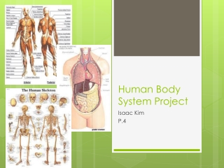
The Visual System: The Structure of the Visual System
460 likes | 785 Views
The Visual System: The Structure of the Visual System. Cornea. The clear bulge on the front of the eyeball Begins to focus the light by bending it toward a central focal point Protects the eye. Parts of the Eye – Cornea. Iris.

The Visual System: The Structure of the Visual System
E N D
Presentation Transcript
Cornea • The clear bulge on the front of the eyeball • Begins to focus the light by bending it toward a central focal point • Protects the eye
Iris • A ring of muscle tissue that forms the colored portion of the eye; creates a hole in the center of the iris (pupil) • Regulates the size of the pupil by changing its size--allowing more or less light to enter the eye
Pupil • The adjustable opening in the center of the eye that controls the amount of light entering the eye (surrounded by the iris) • In bright conditions the iris expands, making the pupil smaller. • In dark conditions the iris contracts, making the pupil larger.
Lens • A transparent structure behind the pupil; focuses the image on the back of the eye (retina) • Muscles that change the thickness of the lens change how the light is bent thereby focusing the image • Glasses or contacts correct problems in the lens’ ability to focus.
Retina • Light-sensitive surface with cells that convert light energy to nerve impulses • At the back of the eyeball
Receptor Cells • These cells are present in every sensory system to change (transduce) some other form of energy into neural impulses. • In sight they change light into neural impulses the brain can understand. • Visual system has two types of receptor cells – rods and cones
Rods • Visual receptor cells located in the retina • Can only detect black and white • Respond to less light than do cones
Cones • Visual receptor cells located in the retina • Can detect sharp images and color • Need more light than the rods • Many cones are clustered in the fovea.
Fovea • The central focal point of the retina • The spot where vision is best (most detailed)
Optic Nerve • The nerve that carries visual information from the eye to the occipital lobes of the brain
Blind Spot • The point at which the optic nerve travels through the retina to exit the eye • There are no rods and cones at this point, so there is a small blind spot in vision.
The Visual System: Color Vision Module 9: Sensation
Color Vision • There are two theories of color vision: • Trichromatic Theory • Opponent-Process Theory
Trichromatic (three-color) Theory • Theory of color vision that says cones are “tuned” to be sensitive to red, green and blue light • All the colors we see are a combination of these three colors. • Similar to the design of a color TV
How do we see color? • Trichromatic (three color) Theory • three different retinal color receptors • Red green blue
Color Deficient Vision • People who lack one of the three types of cones • Usually the red or green receptors are missing • Usually referred to as color blindness • In inherited and found more in males
Opponent-Process Theory- Vision from opposing pairs of color receptors- only one “side” ON at a time
Opponent-Process Theory • Sensory receptors in the retina come in pairs: • Red/Green • Yellow/Blue • Black/White • Only one side is “on” at a time
Opponent Process Theory ON” “OFF” redgreen greenred blueyellow yellowblue black white white black
Opponent-Process Theory • If one sensor is stimulated, the other is inhibited • If one sensor is over-stimulated, and fatigues, the paired sensor will be activated, causing an afterimage