Download
1 / 102
2.74k likes | 7.42k Views
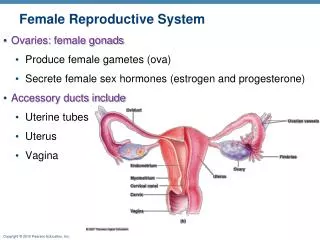
Female Reproductive System. Ovaries: female gonads Produce female gametes (ova) Secrete female sex hormones (estrogen and progesterone) Accessory ducts include Uterine tubes Uterus Vagina. Female Reproductive Anatomy.

E N D
Female Reproductive System • Ovaries: female gonads • Produce female gametes (ova) • Secrete female sex hormones (estrogen and progesterone) • Accessory ducts include • Uterine tubes • Uterus • Vagina
Female Reproductive Anatomy • Internal genitalialocatedinthepelviccavityserveneedes of thereproductivecells and a developing fetus • Ovaries • Uterine tubes (Oviduct) • Uterus • Vagina • External genitalia • The external sex organs
Ovaries • They flank theuterus on eachside • Held in place by several ligaments • Ovarian ligament: anchors ovary medially to the uterus • Suspensory ligament: anchors ovary laterally to the pelvic wall • Mesovarium: suspends the ovary • Thesuspensoryligament and mesovariumare part fo broad ligament: foldwhich „tents” overtheuterus, supports the uterine tubes, uterus, and vagina
Suspensory ligament of ovary Uterine (fallopian) tube Uterine tube Ovarian blood vessels Lumen (cavity) of uterus Fundus of uterus Ampulla Ovary Isthmus Mesosalpinx Infundibulum Mesovarium Broad ligament Fimbriae Mesometrium Round ligament of uterus Ovarian ligament Endometrium Body of uterus Wall of uterus Myometrium Ureter Perimetrium Uterine blood vessels Internal os Isthmus Cervical canal Uterosacral ligament Lateral cervical (cardinal) ligament External os Vagina Lateral fornix Cervix (a) Figure 27.12a
Ovaries • Blood supply: ovarian arteries and the ovarian branch of the uterine artery • Surrounded by a fibrous tunica albuginea • Two poorly defined regions • Cortex: ovarian follicles • Medulla: large blood vessels and nerves
Follicle – tinysaclikestructures, consists of: • Immature egg (oocyte) surrounded by • Follicle cells (one cell layer thick) • Granulosa cells (when more than one layer is present)
Follicles • Several stages of development • Primordial follicle: one layer of a quamouslike follicle cells + oocyte • Primary follicle: single layer of cuboidal or columnar follicle cells + oocyte • Secondary follicle: two or more layers of granulosa cells + oocyte • Late secondary follicle: contains fluid-filled spaces between granulosa cells; coalesces to form a central antrum
Vesicular (Graafian) follicle • Fluid-filled antrum forms; follicle bulges from ovary surface • Ovulation • Ejection of the oocyte from the ripening follicle • Corpus luteum develops from ruptured follicle after ovulation
Tunica albuginea Oocyte Granulosa cells Late secondary follicle Cortex Degenerating corpus luteum (corpus albicans) Mesovarium and blood vessels Germinal epithelium Vesicular (Graafian) follicle Primary follicles Antrum Oocyte Ovarian ligament Zona pellucida Theca folliculi Medulla Ovulated oocyte Corpus luteum Corona radiata Developing corpus luteum (a) Diagrammatic view of an ovary sectioned to reveal the follicles in its interior Figure 27.11a
Female Duct System • Uterine (fallopian) tubes or oviducts • Receivetheovulated oocyte, site for fertilization • Uterus • Vagina
Uterine Tube • Ampulla • Distal expansion with infundibulum near ovary • Usual site of fertilization • Infundibulum an open, funnel-shapedstructurebearingciliated, fingerlikeprojections – fimbriae, stiffen and sweeptheovariansurface, carry an oocyte into uterine tube • Isthmus: constricted region where tube joins uterus
Oocyte is carried along by peristalsis and ciliary action • Theuterinetubecontainssheets of smoothmuscle, and itsthick, highlyfoldedmucosacontains non- and cilatedcells • The oocyte iscarriedtowardtheuterus by a combintionof muscularperistalsis and thebeating of thecilia • Nonciliated cells nourish the oocyte and the sperm Human ovum between fallopopian tube’s fimbriae. Human ovum in the fallopian tube.
Uterus – located in the pelvis • Itis a hollow, muscular organ thatreceives, retain and nourish a fertilizedovum • Body: major portion • Fundus: rounded superior region • Isthmus: narrowed inferior region
Cervix: narrow neck, or outlet; projects into the vagina • Cervical canal communicates with the • Vagina via the external os = mouth • Uterine body via the internal os • Cervical glands secrete mucus that blocks sperm entry except during midcycle
The Uterus is suported laterlly by • Mesometrium—portion of the broad ligament • Lateral cervical (cardinal) ligaments:extend from the cervix and superior part of the vagina to the walls of the pelvis • Uterosacral ligaments secure uterus to the sacrum • Round ligaments bind to the anterior body wall
Uterine Wall • Thewall of uterusiscompose of three layers • Perimetrium: serous layer • Myometrium: interlacing layers of smooth muscle • Endometrium: mucosal lining of theuterinecavity
Suspensory ligament of ovary Uterine (fallopian) tube Uterine tube Ovarian blood vessels Lumen (cavity) of uterus Fundus of uterus Ampulla Ovary Isthmus Mesosalpinx Infundibulum Mesovarium Broad ligament Fimbriae Mesometrium Round ligament of uterus Ovarian ligament Endometrium Body of uterus Wall of uterus Myometrium Ureter Perimetrium Uterine blood vessels Internal os Isthmus Cervical canal Uterosacral ligament Lateral cervical (cardinal) ligament External os Vagina Lateral fornix Cervix (a) Figure 27.12a
Endometrium has two chief strata (layers) • Stratum functionalis(functional layer) • Changes in response to ovarian hormone cycles • Is shed during menstruation • Stratum basalis(deeperbasal layer) • Forms new functionalisafter menstruation • Unresponsive to ovarian hormones
Uterine Vascular Supply • Uterine arteries: arise from internal iliacsinthepelvis, ascendalongthesides of theuterus, and sendbranchesintotheuterinewall • Branches break upintoarcuate arteries: in the myometrium • Thearcuatearteriessendradial branches in the endometrium branch into: • Spiral arteries stratum functionalis • Straight arteries stratum basalis
Lumen of uterus • Thespiral arteriesrepeatedlydegenerate and regenerate, and itistheirspasmsthatcausethefunctionalislayerto be shedduringmenstruation Epithelium Capillaries Uterine glands Venous sinusoids Lamina propria of connective tissue Spiral (coiled) artery Straight artery Endometrial vein Smooth muscle fibers Radial artery Arcuate artery Uterine artery (b) Figure 27.13b
Vagina – a thin wall-tube, 8-10 cm long • Birth canal and organ of copulation • Extends between the bladder and the rectum from the cervix to the exterior
Layers of wall • Fibroelasticadventitia • Smooth muscle muscularis • Stratified squamousmucosa with rugae – stimulatethe penis duringintercourse • In virginsthe mucosa near the vaginal orifice forms an incomplete partition called the hymen(isvascular, maybleedwhenisstretchedorrupturedduring first coitus) • Vaginal fornix: upper end of the vagina surrounding the cervix
Mons pubis Labia majora Prepuce of clitoris Labia minora Clitoris (glans) Urethral orifice Vestibule Hymen (ruptured) Vaginal orifice Anus Opening of the duct of the greater vestibular gland (a) Figure 27.14a
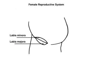
External Genitalia (Vulva or Pudendum): • Thefemalereproductivestructuresthatlieexternal to thevagina: • Mons pubis: fatty area overlying pubic symphysis • Labia majora/largerlips: hair-covered, fatty skin folds – homologue to malescrotum • Labia minora/smallerlips: skin folds lying within labia majora • Vestibule: recess between labia minora – containsexternalopening of theurethraand vagina
External Genitalia • Greater vestibular glands– flankingthevaginalopening • Homologous to the bulbourethral glands • Release mucus into the vestibule for lubricationfacilitatingintercourse
External Genitalia • Clitoris • Composedlargely of erectiletissue – homologous to thepenis • Glans clitoris: exposed portion. Itishooded by a skin fold – calledthepropuce of the clitoris, formed by thejunction of thelabiaminoraflods
Clitoris Labia minora Labia majora Inferior ramus of pubis Pubic symphysis Anus Body of clitoris, containing corpora cavernosa Clitoris (glans) Crus of clitoris Urethral orifice Vaginal orifice Greater vestibular gland Bulb of vestibule (b) Fourchette Figure 27.14b
Duringsexualstimulation engorgewithblood.Help gripthe penis withinvagina, squeezesurethral orfice whichprevents semen and bacteriafromtravelingintothebladderduringintercourse.
Oogenesis • Production of female gametes • Begins in the fetal period • Oogonia (2n ovarian stem cells) multiply by mitosis and store nutrients • Primary oocytes develop in primordial follicles • Primary oocytes begin meiosis but stall in prophase I
Oogenesis • Each month after puberty, a few primary oocytes are activated • One is selected each month to resume meiosis I • Result is two haploid cells • Secondary oocyte • First polar body
Oogenesis • The secondary oocyte arrests in metaphase II and is ovulated!!!!! • If penetrated by sperm the second oocyte completes meiosis II, yielding • Ovum(the functional gamete) • Second polar body
Theformation of gametsintheovaries - oogenesis • Primordial germ cells migrate from the yolk sac to the ovaries and differentiate into oogoniathat divide mitotically. A few, develop into - primary oocytes that enter prophase of meiosis I during fetal development but do not complete that phase until puberty. • After puberty, FSH/LH stimulate the development of several primordial follicles. They start to grow, developing into primary, secondary follicle. • Just before ovulation, the primary oocyte completes meiosis I, producing two haploid cells: first polar body and thesecondary oocyte. • Once a secondary oocyte is formed, it begins meiosis II but then stops in metaphase. The Graafian follicle soon ruptures and releases its secondary oocyte - ovulation. • If sperm are present and one penetrates the secondary oocyte, meiosis II resumes. The secondary oocyte splits into two haploid cells:the ovumand the second polar body. • The nuclei of the sperm cell and the ovum then unite, forming a diploid zygote.
Ovarian Cycle • Monthly series of events associated with the maturationof an egg • Two consecutive phases (in a 28-day cycle) • Follicular phase: period of follicle growth (days 1–14) • Ovulation occurs midcycle • Luteal phase: period of corpus luteum activity (days 14–28) 24-35 days
Follicular Phase • Primordial follicle becomes primary follicle • The primordial follicle is activated • Squamouslike cells become cuboidal • Follicle enlarges to become a primary (1) follicle Theca folliculi 4 3 2 Primary oocyte 1 Zona pellucida Antrum Secondary oocyte 5 Primordial follicles 1 6 8 Secondary oocyte Corona radiata 7 Figure 27.18 (1 of 7)
Theca folliculi 4 3 2 Primary oocyte 1 Zona pellucida Antrum Secondary oocyte 5 Primary follicle 2 6 8 Secondary oocyte Corona radiata 7 Figure 27.18 (2 of 7)
Primary follicle becomes a secondary follicle • Follicularcellsproliferate forming a stratified epithelium (granulosa cells - morethan one cellslayer) around oocyte • Granulosa cells and oocyte guide one another’s development Theca folliculi 4 3 2 Primary oocyte 1 Zona pellucida Antrum Secondary oocyte 5 Secondary follicle 3 6 8 Secondary oocyte Corona radiata 7 Figure 27.18 (3 of 7)
Secondary follicle becomes a late secondary follicle • Connective tissue condensesaroundthefollicle, forming theca folliculi. Granulosa cells with thecal cooperate to produce estrogens – theinner thecal cellsproduceandrogens – granulosa cellsconvert to estrogens • Zona pellucida forms around the oocyte • Fluid begins to accumulatebetweengranulosa cell Theca folliculi 4 3 2 Primary oocyte 1 Zona pellucida Antrum Secondary oocyte 5 6 8 Secondary oocyte Late secondary follicle 4 Corona radiata 7 Figure 27.18 (4 of 7)
Late secondary follicle becomes a vesicular follicle • Antrum forms and expands to isolate the oocyte with itssurroundingcells of granulosa – corona radiata • Vesicular follicle bulges from the external surface of the ovary Theca folliculi 4 3 2 Primary oocyte 1 Zona pellucida Antrum Secondary oocyte Mature vesicular follicle carries out meiosis I; ready to be ovulated 2.5 cm in diameter 5 5 6 8 Secondary oocyte Corona radiata 7 Figure 27.18 (5 of 7)
Ovary wall ruptures and expels the secondary oocyte with its corona radiata - OVULATION • Womenexperience a twinge of pain sometimes • 1–2% of ovulations release more than one secondary oocyte, which, if fertilized, results in fraternal twins (nonidentical) • Identicaltwinsresultsfromfertilization of a single oocyte by a single sperm, followed by separation of thefertilizedegg’sdaughtercellsinearly development Theca folliculi 4 3 2 Primary oocyte 1 Zona pellucida Antrum Secondary oocyte 5 Follicle ruptures; secondary oocyte ovulated 6 6 8 Secondary oocyte Corona radiata 7 Figure 27.18 (6 of 7)
Luteal Phase • Ruptured follicle collapses • Granulosa cells and internal thecal cells form corpus luteum • Corpus luteum secretes progesterone and estrogen
Luteal Phase • If no pregnancy, the corpus luteum degenerates into a corpus albicans in 10 days • If pregnancy occurs, corpus luteum produces hormones until the placenta takes over at about 3 months Theca folliculi 4 3 2 Primary oocyte 1 Zona pellucida Antrum Secondary oocyte 5 Corpus luteum (forms from ruptured follicle) 7 6 8 Secondary oocyte Corona radiata 7 Figure 27.18 (7 of 7)
Establishing the Ovarian Cycle • During childhood, ovaries grow and secrete small amounts of estrogens that inhibit the hypothalamic release of GnRH • As puberty nears, GnRH is released; FSH and LH are released by the pituitary, and act on the ovaries • These events continue until an adult cyclic pattern is achieved and menarche occurs
Establishing the Ovarian Cycle • During childhood, until puberty • Ovaries secrete small amounts of estrogens • Estrogen inhibits release of GnRH
Establishing the Ovarian Cycle • At puberty • Theoneset of pubertyislinked to adiposity • Ifbloodlevels of leptinislow, pubertyisdelayed • Leptin from adipose tissue decreases the estrogen inhibition • GnRH, FSH, and LH are released • In about four years, an adult cyclic pattern is achieved and menarche occurs
Figure 27.19 Feedback interactions in the regulation of ovarian function. Hypothalamus GnRH Travels via portal blood 1 Anterior pituitary 1 FSH LH Early and midfollicular phases
Slide 3 Figure 27.19 Feedback interactions in the regulation of ovarian function. Hypothalamus GnRH Travels via portal blood 1 Anterior pituitary 1 FSH LH 2 2 Thecal cells Granulosa cells Androgens Mature follicle Convert androgens to estrogens Inhibin 2 Early and midfollicular phases
Slide 4 Figure 27.19 Feedback interactions in the regulation of ovarian function. Hypothalamus GnRH Travels via portal blood 1 Anterior pituitary 1 FSH LH 2 2 Slightly elevated estrogen and rising inhibin levels. 3 Thecal cells Granulosa cells Androgens Mature follicle Convert androgens to estrogens Inhibin 2 Early and midfollicular phases