Download
1 / 37
370 likes | 420 Views
بسم الله الرحمن الرحيم. ﴿و ما أوتيتم من العلم إلا قليلا﴾. صدق الله العظيم الاسراء اية 58. Kidney. By Dr. Abdel Aziz M. Hussein Lecturer of Medical Physiology. The role of the kidney in homeostasis may include which of the following :

E N D
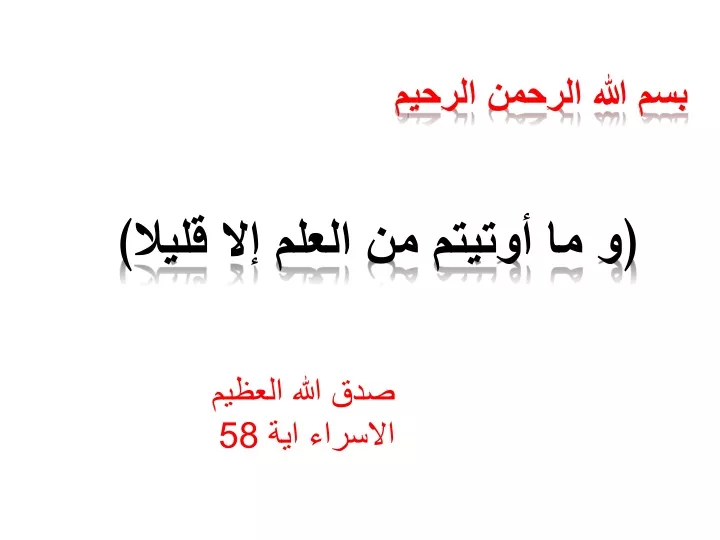
بسم الله الرحمن الرحيم ﴿و ما أوتيتم من العلم إلا قليلا﴾ صدق الله العظيم الاسراء اية 58
Kidney By Dr. Abdel Aziz M. Hussein Lecturer of Medical Physiology
The role of the kidney in homeostasis may include which of the following : • a) Regulation of extracellular fluid composition • b) Regulation of red blood cell formation. • c) Secretion of certain hormones , such as angiotensin II, prostaglandins and kinins • d) Secretion of erythropoietin
Juxta-glomerular Apparatus Tubular pole Vascular pole Def.,: It is a combination of vascular, tubular and interstitial cells Site: Vascular pole of Bowman's capsule
Juxta-glomerular Apparatus Components:
Macula Densa Function • Monitor NaCl concentration in DCT (stimulated by low NaCl) Afferent ↓ GFR ↑ GFR V.C. Efferent Angiotensin II
Juxta-glomerular cell Function • A) Synthesis, store and release of renin • B) acts as Baroreceptors (detect tension in wall of afferent arterioles) JG cells ↓ wall tension ↓ Renal Blood Flow Renin
Extra-glomerular Mesangial cell or Lacis (Polkisson) Function • Form functional syncitium with macula densa and JG cells
Functions of JGA • The only function of JGA is synthesis and secretion of renin Actions of Renin: Stimulates Na reabsorption from PT Stimulates Na and H2O reabsorption from intestine Stimulates ACTH, ADH, etc…. V.C. Thirst and salt appetite Increase force of myocardial contraction Angiotensin II • Small dose (V.C. of efferent) • Large dose (generalized V.C.) ACE Aldosterone Angiotensin I Renin Angiotensinogen
Control of Renin Secretion A) Stimuli for Renin Release: they are 3 stimuli
1. Mechanical stimuli in the form of decreased wall tension 3. Nervous stimuli via B1 receptor 2. Chemical stimuli as in Macula densa mechanism
Macula Densa Mechanism 1. Decrease of GFR 2. Decrease flow through renal tubules 3. Decrease NaCl at Macula densa 4. Macula densa secrete PGI2 5. PGI2 stimulate JG cells to secrete renin angiotensin II → V.C. of efferent arteriole → ↑ GFR
Macula Densa Mechanism 1. Increase of GFR 2. Increase flow through renal tubules 3. Increase NaCl at Macula densa 4. Macula densa secrete adenosine 5. Adenosine V.C. of afferent arterioles ↓GFR
Decreased Wall Tension Renin Decreased Ca → increase cAMP Decreased wall tension Decrease of ABP ↓ Renal Blood Flow
Increased Sympathetic activity Decrease of renin Decrease of ABP
Control of Renin Secretion B) Inhibitors of Renin: include • Angiotensin II • Angiotensin III • Atrial natriuretic peptide (ANP) • ADH • Hyperkalemia • Hypernatremia Do not forget: 4 A 2 H
Renal O2 Consumption • Is characterized by; • 1) 8% of total O2 consumption i.e. 18- 20 ml /min for both kidneys or 6 ml/min/100 gm kidney tissue • 2) Arteriovenous O difference is low i.e. 1.5 ml O2/ 100 ml blood compared to 4 -5 ml /100 ml in other tissues 19 ml % 17.5 ml %
Renal O2 Consumption • 3) Renal O2 consumption is a flow dependent i.e. increase of RBF → increase of renal O2 consumption • 4) Most of O2 consumption is used for tubular reabsorption especially Na Increase of RBF Increase of renal O2 consumption
Renal Blood Flow Def.: • It is the fraction of CO that supplies both kidneys i.e. renal fraction RBF
Renal Blood Flow Value: • ¼ CO or 1200ml/min or 4 ml/ 1 gm kidney tissues Significance: • Is high to ensure high GFR NOT to supply excess O2 for excess metabolism
Renal Blood Flow Distribution: A) 10% supply non-functioning structures of kidney • a) capsule • b) pelvis • c) perinephric fats B) 90% functioning structures • Cortex → 88% - 89% • Medulla → 1- 2%
Renal Blood Flow 4-5 ml/ min 1 gm Cortex 0.7- 1 ml /min/ 1gm Outer Medulla 0.2 – 0.25 ml /min/ 1gm Inner Medulla
Low Medullary RBF High length of vasa recta High viscosity of medullary blood flow Small number of vessels