Brain function – the evidence from brain damage
270 likes | 442 Views
Brain function – the evidence from brain damage. Brain damage to a specific area and you cant talk? Maybe that area controlled speech?. Language Centres – Left Hemisphere. Broca’s Area. Broca’s Area Located in the frontal lobe of the LEFT hemisphere only

Brain function – the evidence from brain damage
E N D
Presentation Transcript
Brain function – the evidence from brain damage Brain damage to a specific area and you cant talk? Maybe that area controlled speech?
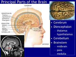
Broca’s Area • Broca’s Area • Located in the frontal lobe of the LEFT hemisphere only • Production of articulate speech, clear and fluent • Moves the muscles that are required to speak • Involved in analysing the grammatical structure of sentences, helps us extract meaning from language
Broca’s Aphasia • Damage to broca’s area • Broca’s aphasia (non fluent aphasia) = Trouble with speech production, speech consists of very short and simple sentences, mainly verbs and nouns • Reading and writing not as effected (can be in some cases though)
Test for Broca’s Aphasia ‘stool, is it boy, is it that landing down, girl is laughing, and cookie jar……….ok um…..window….curtains and out the garden and trees low grass and um lady washing the dishes and hot and cold water….plashing running, floor , and the two cups of a, two cups of ah, two cups of ah, coffee or um….empty um……..cupboard and cupboards …..um……washing…..flashing and um earth to roof’
Wernicke’s Area • Located in the temporal lobe of the left hemisphere only, next to the primary auditory cortex • Involved in the Interpretation of speech, referred to as the language comprehension center • Vital for locating appropriate words from memory to express meaning
Wernicke’s Aphasia • Damage to Wernike’s area • Wernicke’s aphasia (fluent aphasia) = Trouble with speech comprehension. Also cant produce meaningful sentences, can string words together but what they say is nonsensical.
Test for Wernicke’s Aphasia ‘Mother is away here working her work to get her better, but when she’s looking the two boys looking in the other part. She’s working another time.’
Neglect Syndrome Nothing on you left! Damage to the right!
Neglect syndrome • Most commonly associated with damage to the right parietal lobe • Results in the patient completely ignoring the left side of their world, even the left side of their body. • Patients eat only the food on the right side of their plate, shave the right side of their face, wash the right side of their body etc. • A problem of attention, not blindness!
Split Brain Surgery • Severing the Corpus Callosum • Used to treat severe epilepsy • Disables communication between the left and right hemispheres
Information from left visual field is sent to the right half of each eye and then processed in the right hemisphere. • Information from the right visual field is sent to the left half of each eye and then processed inthe left hemisphere • Information from the central visual field is processes in both hemispheres
Split Brain Experiment – Roger Sperry and Michael Gazzaniga • Object presented to left visual field (processed in right hemisphere) • Cannot name object • Can pick correct object up • Object presented to right visual field (processed in left hemisphere) • Can name object • Conclusion – language processing occurs on LEFT and communication disrupted due to severed corpus callosum
http://www.youtube.com/watch?v=aCv4K5aStdU 4 mins • http://www.youtube.com/watch?v=lfGwsAdS9Dc&feature=related 10 mins
2006 Exam Question ANSWER Tony is looking straight ahead at a projector screen. His right eye is covered with a blindfold. An image of a penguin is projected onto the middle of the screen. This information would be registered in _______________ hemisphere(s). • A. the right • B. the left • C. both the left and right • D. neither the left nor right • C = An image in the centre of the field of view will be processed in both hemispheres because the image will fall on the left and right sides of the retina in each eye.
2007 Exam Question ANSWER Visual information received by the right eye is processed in the • A. parietal lobe of the left hemisphere only. • B. occipital lobe of the left hemisphere only. • C. parietal lobe of right and left hemispheres. • D. occipital lobe of right and left hemispheres. • D = Images falling on the left side of each retina are processed in the left occipital lobe and images falling on the right side of each retina are processed in the right occipital lobe. Almost half of the students chose the incorrect alternative B. • This question was similar to Question 6 on the 2006 paper, which was also poorly answered.
2010 Exam Question ANSWER Visual images received in the left visual field are processed in the • A. occipital lobe of the left and right hemispheres. • B. temporal lobe of the right hemisphere only. • C. occipital lobe of the right hemisphere only. • D. occipital lobe of the left hemisphere only. Option A (occipital lobe of the left and right hemispheres) would only be correct for images in the central visual field. The correct answer is C
Perceptual Anomalies Are you sure what you see is real?
Motion after effect • Perceptual illusion of the movement of a stationary object following exposure to motion • Cause is not fully understood • Seems clear that eye movement and neurons in the visual cortex are involved
Change Blindness • Difficulty in observing large changes to visual scenes • The change must take place during a brief visual disruption • Occurs when change is expected and unexpected. • Attention clearly plays a role as well as short term memory.
Inattentional Blindness • Failure to notice a change in a scene when the same scene continually remains in sight • Not the same as change blindness • No disruption • EG. Gorilla experiment
http://www.youtube.com/watch?v=mAnKvo-fPs0 • http://www.youtube.com/watch?v=ubNF9QNEQLA
Synaesthesia • Stimulation of one sense produced additional and unusual experiences in another sense. • 5 senses: Touch, sound, smell, taste, sight. • Grapheme synaesthesia (colour synaesthesia): The number 3 will always appear as blue regardless of its actual colour. • It is involuntary • Effects roughly 1 in 2000 • Unclear as to why it occurs – possibly super sensitive receptors or neural wiring disrupted.