Download
1 / 54
570 likes | 677 Views
Explore real-life cardiology cases in dogs with practical ECG interpretations and treatment strategies. Follow Jake, a Boxer with dilated cardiomyopathy and congestive heart failure, through diagnosis, therapeutic plans, and follow-ups.

E N D
Practical CardiologyECG Case Studies Wendy Blount, DVM Nacogdoches TX
http://www.wendyblount.com • Go to the website • Click on “Presentation Notes” either on the left or at the bottom of the page • Choose the presentation you want • Download all materials
http://www.wendyblount.com • Treatment by Arrhythmia • Antiarrhythmic Drug Classes and Doses • Arrhythmia Description and Classification • This PowerPoint
Jake Signalment • 9 year old male Boxer Chief Complaint • Deep cough when walking in the morning, for about one week • Appetite is good
Jake Exam • Weight 81.9 – has lost 5 pounds in 3 months (BCS 3) • Temp 101.4 • Mucous membranes pink, CRT 3.5 seconds • Subtle dependent edema on the lower legs • Jugular veins distended • Harsh lung sounds • 3/6 holosystolic murmur, PMI left apex • Heart rate 160 per minute • Respirations 55 per minute • Femoral pulses somewhat weak
Jake Differential Diagnosis - Cough • Respiratory Disease • Cardiovascular Disease • Both Diagnostic Plan (B Client) • Blood Pressure • 150 mm Hg systolic (Doppler) • Chest x-rays
Jake Diagnostic Plan (B Client) • Chest X-rays • Massively enlarged heart (VHS 12.5) • Enlarged LA, LV (dorsally elevated trachea) • Enlarged pulmonary veins • Perihilar pulmonary edema • Left congestive heart failure
Jake Immediate Therapeutic Plan (10 am) • Furosemide • 80 mg IM • 4 hours later • Respiratory rate is 36 per minute
Jake Diagnostic Plan – 2nd Wave (2 pm) • EKG • Normal Sinus Rhythm • Echocardiogram (video) • Enlarged LV, myocardium is hardly moving • IVS bowed to the right due to LV dilation • Measurements confirm LV enlargement, LA enlargement and myocardial failure • EF 15% • FS 7% • LA:Ao 2.1 Diagnoses: Dilated Cardiomyopathy with biventricular CHF
Jake – Dx & Tx Recommendations • Congestive Heart Failure • CBC, serum panel and electrolytes • Furosemide 80 mg PO BID • Enalapril 20 mg PO BID • Recheck mini-panel and electrolytes in 3-5 days • Recheck chest rads and BP 3-5 days • Dilated Cardiomyopathy • Thyroid panel (TSH, T4, FreeT4) • Pimobendan 10 mg PO BID (declined) • Carnitine 2 g PO BID • Recheck echo, chest rads, BP, EKG, mini-panel/lytes 60 days (sooner if respiratory rate >40 at rest)
Jake - Bloodwork Carnitine for DCM • Boxers with genetic defect need extra carnitine • Plasma levels have low sensitivity • Myocardial biopsy is usually required CBC, Mini-panel - BUN, creat, glucose, TP, SAP, ALT • Normal Electrolytes, Thyroid panel • Not done
Jake – Follow-Up Recheck – 6 days • BUN 30 (n 10-29) • Creat normal • Electrolytes not done • Chest x-rays not done No additional rechecks were done, owner did not monitor respiratory rate at home
Jake – Follow-Up 4 months later… • Chief complaint – • Doing well until last week • poor energy, coughing again, not eating • Heart sounds (audio file) • Chaotic heart sounds with pulse deficits on auscultation • “tennis shoes in a dryer”
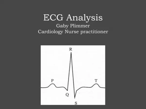
Interpreting the ECG • Heart Rate • Rhythm • Normal Sinus Rhythm • Similar P QRS and T for each beat • Regular heart rate • Respiratory Sinus Arrhythmia • Similar P QRS and T for each beat • Heart rate increases with inspiration & decreases with expiration • Arrhythmia • P wave - width and height • PR interval - length • QRS - width and height
Jake – Follow-Up 25 mm/sec • “Bic Pen x 10” • At 25 mm/sec, 150 mm of ECG = 6 seconds • A Bic Pen is 150 mm long • So the number of QRS complexes in a Bic Pen x 10 = heart rate
Jake – Follow-Up 25 mm/sec • Heart Rate • 200 bpm (tachycardia) • Rhythm (NSR, RSA or arrhythmia) • irregularly irregular - arrhythmia
Jake – Follow-Up • P wave • (normal 1 box wide x 4 boxes tall) • not present • PR interval (normal 1.5-3.25 boxes) • no P wave – can’t measure • QRS • (normal 1.5 boxes wide x 30 boxes tall) • 2 boxes wide x 26 boxes tall • Wide QRS = LV enlargement 25 mm/sec Diagnosis – Atrial Fibrillation
Jake – Treatment • Recommended treatment • Pimobendan for DCM (declined before) • Digitalis for Afib • Treatment was declined, and Jake was euthanatized 1 week later • Most dogs with DCM are gone within 3 months of becoming symptomatic, if treated with furosemide & ACE. • Survival is likely much shorter – days to weeks – if untreated. • Adding Pimobendan increases mean survival to 130 days. • Median survival for dogs with DCM and Afib is 3 weeks, without Pimobendan
Dilated Cardiomyopathy Common ECG Findings • Wide P wave • LA enlargement • Tall R wave • LV enlargement • Atrial fibrillation • VPCs • Ventricular arrhythmias
Atrial Fibrillation Why Treat?? • Heart rate around 250 beats per minute • Myocardial failure will result within 3-6 weeks • Ventricles can not fill properly – forward heart failure Treatment • Conversion would be ideal • But this is not easy to accomplish in very sick hearts • Can attempt in big dogs with normal hearts and primary Afib, not dogs with DCM • Can try medical conversion with quinidine • Or Anesthesia and conversion with electric shock
Atrial Fibrillation Treatment – Afib in unhealthy hearts • Slow the heart rate at the AV node (goal 150 bpm) • Digoxin • Weak positive inotrope • Beta blockers • Negative inotrope – probably contraindicated if DCM • Calcium channel blockers • Diltiazem SR (Plumb dose) DON’T USE BETA BLOCKER AND CALCIUM CHANNEL BLOCKER TOGETHER!!
Tom 5 year old neutered male DSH Chief Complaint • Outdoor cat, owners think he was hit by a car • Tom is laterally recumbent, and breathing hard Exam • T 96.5, P- 100, R – 66 • No evidence of trauma
Tom ECG 1 • Heart Rate - 120 • Rhythm – regular • no P waves • QRS – deep S wave, wide, bizarre QRS Dx – atrial standstill, L ventricular escape rhythm
Tom i-STAT EC8+ • K 10.9 mEq/L, iCa++ 0.96 mmol/L • pH 7.08, HCO3 11 mEq/L • Grapefruit sized very firm bladder
Tom Treatment • Place indwelling urinary catheter & IV catheter • Begin 0.9% NaCl at 15 ml/hr • 1 unit regular insulin IV • 5cc 50% dextrose diluted in 15 cc fluids, given over 1 hour; added 5%dextrose to fluids ECG 2 – 6 minutes later
Tom • ECG 2 – 6 minutes later
Tom ECG 2 – 6 minutes later • Heart rate 140 • No P waves • QRS less abnormal • T wave not as tall
Tom ECG 3 – 1 hour after presentation • Heart rate 120 • No change for the past 45 minutes Treatment • Ca-gluconate 2cc IV slowly over 20 minutes
Tom ECG 4 – 2 hours after presentation – T 98.9 • Heart rate 120, normal sinus rhythm • P waves have returned, but wide and inverted • QRS and T normal
Tom ECG 5 – 5 hours after presentation • Heart rate 130 • Normal sinus rhythm • P waves have returned to normal
Tom Follow-up i-STAT EC8+ • iCa++ normal, K 6.6 mEq/L • HCO3-- 16.3 mEq/L, pH 7.29 Tom began eating the next day, the urinary catheter was removed, and he was discharged 2 days later. • He was azotemic on presentation, but this resolved with treatment
Gabby 6 month female DSH Presented for OHE Exam - HR 100 • No other abnormal findings • Preanesthetic bloodwork normal
Gabby Pre-Anesthetic ECG • Heart rate • P rate is 160 bpm, QRS rate is 100 bpm • Rhythm • no consistent PR interval • P and QRS complexes are disassociated, but each regular 20mm = 1 mV 25 mm/sec 3rd Degree AV block
3rd degree AV block 3rd Degree AV block is the most common cause of bradycardia in the cat Treatment- cats • Often no treatment needed for cats • AV node pacemaker is 100 per minute • AV node pacemaker is 40-60 per minute in the dog • Cats do well unless they undergo anesthesia • Avoid drugs that increase vagal tone • Alpha blockers – Dexdomitor, Rompun
Gabby • Gabby was not spayed at 6 months of age • When she reached 7 years of age, she had her 4th litter • She was referred to Drs. Miller and Gordon at TAMU for spay • When induced, her heart rate immediately fell to 40 and was progressively dropping • A temporary pacemaker was placed • Gabby was spayed and recovered uneventfully • Gabby turned 17 years old this year
Gabby Dear Doc, Because you took away my favorite pastime, I have turned to a life of substance abuse. It’s your fault. Love, Gabby
3rd degree AV block in Dogs • Usually presents for syncope • “Cannon wave” jugular pulses (bradycardia) • Treated with pacemaker implantation • Drug therapy not usually successful • Usually no response to atropine • Atropine often makes 2nd degree block go away • Some have tried theophylline • Prognosis poor without pacemaker • If lactate is high, emergency pacemaker is needed
3rd degree AV block in Dogs Pre-Operative ECG • Atrial rate = 200 per minute • Ventricular rate = 40 per minute 50 mm/sec
3rd degree AV block in Dogs Post-Operative ECG • Ventricular rate = 100 50 mm/sec
Susie Signalment • 12 year old spayed miniature schnauzer Chief Complaint • Episodes of Confusion Exam • G3 dental tartar • Alternating periods of normal heart rate, tachycardia and bradycardia • Pulse deficits during tachycardia
Susie Work-up • CBC, panel, electrolytes, UA normal • Chest x-rays
Susie Work-up • CBC, panel, electrolytes, UA normal • Chest x-rays Vertebral Heart Size = 10.7 (normal 8.5-10.5) Enlarged main pulmonary artery
Susie Work-up • CBC, panel, electrolytes, UA normal • Chest x-rays • Susie is not on heartworm prevention
Susie ECG • Heart Rate • Very erratic an impossible to estimate • >200 bpm for periods of up to 2-4 seconds • Some periods of normal heart rate • Periods of asystole for up to 2-4 seconds 25 mm/sec
Susie ECG • Rhythm – arrhythmia • P wave (normal 1 box wide x 4 boxes tall) • Some P waves missing and some inverted • Wandering pacemaker, failure of pacemaker and acceleration of pacemaker in the SA node 25 mm/sec