Download
1 / 54
550 likes | 683 Views
Light Microscopy and Electronic Imaging for the Biomedical Sciences. E. D. Salmon and Kerry Bloom Biology 188. History of the Microscope, Thomas E. Jones. http://www.utmem.edu/~thjones/hist/hist_mic.htm. See also Molecular Expressions, a Microscope Primer at:

E N D
Light Microscopy and Electronic Imaging for the Biomedical Sciences E. D. Salmon and Kerry Bloom Biology 188
History of the Microscope, Thomas E. Jones http://www.utmem.edu/~thjones/hist/hist_mic.htm See also Molecular Expressions, a Microscope Primer at: http://micro.magnet.fsu.edu/primer/index.html There is one very early description of an isolated use of spectacles. Pliny the Elder wrote the following in 23-79 A.D.: "Emeralds are usually concave so that they may concentrate the visual rays. The Emperor Nero used to watch in an Emerald the gladatorial combats." The modern reinvention of spectacles occurred around 1280-1285 in Florence, Italy
“Microscope” Named and a 2-lens "Huygens Eyepiece Introduced in Early 1600’s Italian microscope Galileo might have used
Hooke Microscope Had a Resolution of About 5 mm “Cells” Discovered
Leeuwenhoek's Secret Lenses: Leeuwenhoek's method of making the tiny, high-quality and high power lenses was kept secret. A study has recently been done on the few remaining copies of Leeuwenhoek's microscopes, and it appears that some of the lenses may have been made by grinding, while the best ones were blown. Leeuwenhoek learned that when a glass bulb is blown, a small drop of thickened glass forms at the bottom of the bulb (much like a drop sits in the bottom of a blown soap bubble.) By carefully breaking away the excess glass, this tiny drop can be used as a lens.
Chromatic and Spherical Aberration Limited Resolution While the 18th century produced some great mechanical improvements for the microscope, making it much more sturdy and easy to use, the images obtainable remained rather blurry with colorful halos around objects. This was largely due to the problems of "Chromatic and Aspheric Aberration." The reason the single lens "simple" microscopes remained important throughout the century was that a single lens system has much less aberration because the distortion becomes synergistic with multiple lenses. This allowed simple microscopes to attain around 2 micron resolution, while the best compound microscopes were limited to around 5 microns.
Chromatic Aberration Corrected by the Achromatic Doublet Chester More Hall Makes the Discovery in 1730, diddles, and John Dolland Learns the Secret, and Patents it in about 1759.
Spherical Aberration Not Solved Until 1830 by Joseph Jackson Lester Tulley/Lister Corrected Lens Microscope, 1830's Adjustable Objective by Ross, circa 1840
Abbe Discovers in 1877 The Importance of Numerical Aperture (NA = nsinq) for Resolution Developed Apochromatic Optics
Köhler illumination was first introduced in 1893 by August Köhler of the Carl Zeiss corporation as a method of providing the optimum specimen illumination
Objective Turrets Developed and Modern Condenser Design Parfocal Objectives Abbe condensers with Cond. Diaphragm and Turret
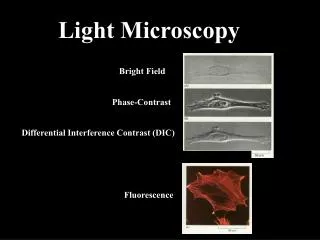
Phase Contrast Gives Contrast to Structural Detail in Transparent Specimens In focus Image: Get phase contrast by slight out-of-focus, but loss of resolution
Differential Interference Microscopy (DIC) Invented by Nomarski and Smith in 1960’s
Mono-Clonal and Affnitiy Purified Antibody Methods and Beginning of Molecular Probe Development Began in 1970’s Multi-Wavelength Fluorescence Microscopy: Co-Localization of Different Molecules Relative To Cellular Structures
Video-Enhanced Contrast Methods Developed in Early 1980’s by Inoue and Allen Revealed Cellular Structures and Macromolecular Complexes Invisible by Eye or Film
Video-Enhanced DIC Microscope System from 1985
VE-DIC Motility Assays Lead to Discovery of Microtubule Motor Proteins Like Kinesin in Mid-1980’s and After
8 nm Step 5 pN Stall Force 100 Steps/sec at No Load 1 ATP Hydrolized Per Step Optical Trap Coverslip
Simulation from Ron Milligan and Ron Vale of Kinesin Mechanochemical cycle
Fluorescence microscopy pushed forward in early 1980’s by new fluorophores (start of Molecular Probes) and intensified video cameras • Detect fluorescence invisible to eye or film • Quantitative fluorescence measurements • Fluorescent protein analogs of live cells • Ratio measurements for ion dynamics (e.g. Fura 2 for calcium ion…) • Molecular dynamics from Measurements of fluorescence recovery after photobleaching (FRAP)
Today: e.g. Hamamatsu Orca ER Cooled CCD Camera • Low readout noise (~8 electrons) • High Quantum Efficiency • Broad spectral response • Fast readout: ~8MHz • No distortion • 1024x1024 pixels • >20,000 e deep wells
Measurements of Fluorescence Recovery After Photobleaching (FRAP) Shows that Alexa488- or GFP-Mad2 Turns-Over Rapidly at Unattached Kinetochores ( a 20-25 sec half-life) Howell et al., 2000, J. Cell Biol. 150:1-17.
1987: John White and Brad Amos Invented Modern Laser Scanning Confocal Fluorescence Microscope
In Mid 1990’s Went from Single Photon to Multiphoton Imaging
The Modern Era of Light Microscopy • New microscope optics generate brilliant images over wide wavelengths • Computers control x-y-&z specimen position, wavelength selection, illumination and image acquisition • Electronic cameras quantitatively record light intensity of specimens invisible or undetectable by eye or film • Confocal and deconvolution methods give 3-D views of cellular architectural dynamics • New fluorescent molecular probes and biophysical methods report on the temporal and spatial activities of the molecular machinery of living cells and single molecule imaging • Micromanipulation, ablation, force measurement
Modern Upright Research Light Microscope (1995) *Bright, High Contrast Optics *Epi-Fluorescence *Phase-Contrast *Polarization *DIC *Diffraction Limited Resolution *Multiple Ports *Auto. Photography *Electronic Imaging- (Video---CCD)
The Modern Era of Light Microscopy • New microscope optics generate brilliant images over wide wavelengths • Computers control x-y-&z specimen position, wavelength selection, illumination and image acquisition • Electronic cameras quantitatively record light intensity of specimens invisible or undetectable by eye or film • Confocal and deconvolution methods give 3-D views of cellular architectural dynamics • New fluorescent molecular probes and biophysical methods report on the temporal and spatial activities of the molecular machinery of living cells and single molecule imaging • Micromanipulation, ablation, force measurement
In early 1990’s, went to semi-automated, multimode,wide-field microscopes with cooled CCD cameras, shutters, filter wheels and computer control
Multi-Wavelength Immunofluorescence Microscopy
Confocal Scanning Head Nikon TE300 inverted microscope Filter Wheel Orca ER CCD PC with MetaMorph software Laser Input (fiber optic) Focus motor
High Resolution, High Signal-Noise, 1Kx1K Pixel Images Recorded in 200ms Immunofluorescence Microscopy of Microtubules (Green) And Chromosomes (Red) In Mitotic PtK1 Cell
Molecular Fluorescent Probes • Specific Fluorescent Dyes (e.g. DAPI) • Covalently bind fluorescent dye to purified protein • Fluorescent Antibodies (e.g immunofluorescent labeling with primary and fluorescent secondary antibodies) • Express in cells Green Fluorescent Protein (GFP) fused to protein of interest
Cellular Imaging is Key to Understanding Protein Function in Cells Genomics Cellular Imaging e.g. GFP-Fusion Proteins Proteomics
Alexa-488-Eb1 Bound to the Growing Ends(10 mm/min)of Microtubulesin Early PrometaphaseSpindle in Xenopus EggExtracts(Jen Ternauer)
Cdc20 Persists At Kinetochores Throughout Mitosis and Exhibits Fast Kinetics: FRAP t1/2 = [4 sec (attached) 25 sec (unattached] Green: GFP-Cdc20 At Kinetochores Red: Phase Contrast Images of PtK1 Tissue Cells
Biological System: Budding Yeast • Saccharomyces cerevisiae • Short cell cycle. • Genetics. • Ease of Gfp constructs. • Conserved mitotic processes.