Download
1 / 40
420 likes | 637 Views
The Neuroplasticity Hypothesis of Alzheimer's Disease A Bio-Psycho-Social Systems Theory and Neural-Network Perspective for Integrating the Alzheimer Field and Providing an Approach to Treatment and Prevention. J. Wesson Ashford, M.D., Ph.D.

E N D
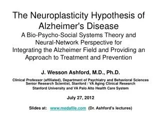
The Neuroplasticity Hypothesis of Alzheimer's DiseaseA Bio-Psycho-Social Systems Theory and Neural-Network Perspective for Integrating the Alzheimer Field and Providing an Approach to Treatment and Prevention J. Wesson Ashford, M.D., Ph.D. Clinical Professor (affiliated), Department of Psychiatry and Behavioral Sciences Senior Research Scientist, Stanford / VA Aging Clinical Research Stanford University and VA Palo Alto Health Care System July 27, 2012 Slides at: www.medafile.com(Dr. Ashford’s lectures)
NEUROPLASTICITY HYPOTHESIS OF ALZHEIMER PATHOLOGY • AD ATTACKS NEUROPLASTIC MECHANISMS OF THE BRAIN • SENILE PLAQUES AND NEUROFIBRILLARY TANGLES ARE THE VISIBLE SCARS OF THE ATTACK, NOT THE PATHOGENIC AGENTS • THERE IS NO EVIDENCE THAT THE BETA-AMYLOID PROTEIN IS HARMFUL TO THE BRAIN OR CORRELATED WITH THE DEMENTIA • NEUROFIBRILLARY TANGLES ARE CORRELATED WITH THE DEMENTIA, BUT NOT CAUSAL OF DEMENTIA • CAUSAL GENETIC FACTORS CAN ALL BE RELATED TO THE AMYLOID PRE-PROTEIN AND EITHER ITS OVER-PRODUCTION, MODULATION OF BETA- AND GAMMA-SECRETASE CLEAVAGE, OR CLEARANCE • THE HARMFUL EVENT IS THE HYPER-PHOSPHORYLATION OF THE MICROTUBULE-ASSOCIATED PROTEIN TAU, LEADING TO PAIRED HELICAL FILAMENTS, NEUROPIL THREATS, AND LOSS OF SYNAPSES • THE ALZHEIMER ATTACK IS A MODEL TO UNDERSTAND NEUROPLASTICITY
Dementia Definition • Multiple Cognitive Deficits: • Memory dysfunction • especially new learning, a prominent early symptom • At least one additional cognitive deficit • aphasia, apraxia, agnosia, or executive dysfunction • Cognitive Disturbances: • Sufficiently severe to cause impairment of occupational or social functioning and • Must represent a decline from a previous level of functioning
Alzheimer’s Disease • First described by Alois Alzheimer, a German neuropathologist, in 1906/7 • Observed in a 51-year-old female patient with paranoia, memory loss, disorientation, and hallucinations • Postmortem studies characterized senile plaques and neurofibrillary tangles (NFTs) in the cerebral cortex • Senile plaques: Extracellularaccumulation of insoluble fragments of beta-amyloid (A1-42) • NFTs: Intracellular accumulation of hyperphosphorylated tau strands
Diagnostic Criteria For Dementia Of The Alzheimer Type(DSM-IV, APA, 1994) • Memory Impairment 1. Multiple Cognitive Deficits 2. Other Cognitive Impairment • Deficits Impair Social/Occupational Function • Course Shows Gradual Onset and Decline • Deficits Are Not Due to: 1. Other CNS Conditions 2. Substance Induced Conditions • Do Not Occur Exclusively during Delirium • Not Due to Another Psychiatric Disorder
Alzheimer Pathology and Senile Dementia • In 1968, Blessed, Tomlinson, and Roth showed that counts of neurofibrillary tangles correlated with dementia severity in older individuals • They did not find a correlation between counts of senile plaques and dementia severity (and this relationship has never been firmly established)
Cholinergic Changes in AD - 1976 • The most prominent neurotransmitter abnormalities in AD are cholinergic • Reduced activity of choline acetyltransferase (synthesis of acetylcholine)1 • Reduced number of cholinergic neurons in late AD (particularly in basal forebrain)2 • Selective loss of nicotinic receptor subtypes in hippocampus and cortex1,3 1. Bartus RT et al. Science. 1982;217:408-414. 2. Whitehouse PJ et al. Science. 1982;215:1237-1239. 3. Guan ZZ et al. J Neurochem. 2000;74:237-243.
Cholineric Hypothesis of AD • Anti-muscarinic agents cause memory impairment – similar to AD • Cholinergic agents improve memory function • Acetyl-cholinesterase is decreased in the AD brain • 1976 – 3 studies show decreased choline-acetyltransferase in AD brain • 1981 - Loss of cholinergic neurons in nucleus basalis of Meynert in AD • Cholinergic agents considered for treatment – lecithin, agonists • Cholinesterase inhibitors (AChE ) considered for treatment of AD • 1st double blind study - physostigmine - Ashford et al., 1981 • 1st successful treatment of AD - physostigmine - Thal et al., 1983 • 4 AChEI medications subsequently approved by FDA for treating AD • AChEIs presumably increases acetylcholine at synapses • Improvement in cognition (? 6-12 months better) • Improvement in function (ADLs, variable) • Improvement in behavior (? basal ganglia) • Loss of nicotinic brain receptors is biggest chemical change in AD brain • Slowing of disease course • Treatment delays nursing home placement • There is loss of benefit with delay of treatment • May treat disease process, not just symptoms
Problems with the Cholinergic Hypothesis • Many cholinergic neurons throughout brain, spinal cord, but only discrete groups of ACh neurons are affected in AD • Numerous other neurotransmitter systems are affected in AD • Cholinergic agents are only modestly effective in treating AD, slowing progression • No clear relationship between acetylcholine and microscopic neuropathological features
Specific groups of cholinergic, serotonergic, and noradrenergic that project to the cortex, and glutamatergic and somatostatinergic GABA neurons of discrete cortical regions are selectively affected in Alzheimer’s disease Most affected) by AD -memory-write signal • Cortex • - Glutamate neurons • - highly affected by AD • - detail memory • - Rx: memantine • GABA neurons • - Somatostatinergic • neurons affected by AD • – memory modulation Rx: cholinesterase inhibitors (not affected by AD - movement) (Affected by AD -operant conditioning) (Affected by AD early - Classical conditioning)
Neurotransmitter therapies • Cholinergic system – • Modest clinical benefit of cholinesterase inhibitors • No benefit from any other treatments • No evidence of benefit in early AD or mild cognitive impairment • Serotonergic system – none effective • Noradrenergic system – none effective • Dopaminergic system – none effective • Glutamate system • NMDA modulator memantine • Slight improvement in moderate to severe AD • May improve behavior or make it worse • May increase life-expectancy to near normal • GABA system – none effective
Discrete regions of the cerebral cortex are selectively affected by Alzheimer pathology Brun & Englund, 1986
CORTICAL PATHOLOGY AFFECTS LOCATIONS OF HIGH MEMORY STORAGE Shown on the next slide is a correlation analysis between brain perfusion (SPECT) and dementia severity (transformed from the MMSE) (Ashford et al., 2000). This finding is consistent with observations using numerous other modalities, e.g, PET
Relation of SPECT severity to duration of dementia (years)Shih, Ashford et al., 2000
Alzheimer pathology affects regions of the cortex that have a high capacity and responsibility for memory storage Sensory, Perception, Memory systems of cortex – Ashford, Coburn, Fuster, 1998
COGNITIVE PATHOLOGY REFLECTS FAILURES OF MEMORY STORAGE Shown on the next slide is an “Item Response Theory” analysis of the items of the Mini-Mental State Exam (MMSE). The results show that items which are more involved in memory processing are affected earlier in AD (Ashford et al., 1989, 1995).
Neuroplasticity Hypothesis of Alzheimer’s DiseaseAt all Bio-Psycho-Social System Levels, the Alzheimer Process Attacks Mechanisms Associated with Memory • SOCIAL SYSTEMS REQUIRING MEMORY • INSTRUMENTAL ADLs - EARLY • BASIC ADLs - LATE • PSYCHOLOGICAL MEMORY SYSTEMS • PRIMARY LOSS OF SHORT-TERM MEMORY • LEARNING PROCESSES – CLASSICAL, OPERANT • LATER LOSS OF LEARNED SKILLS • NEURONAL MEMORY SYSTEMS • CORTICAL • GLUTAMATERGIC LARGE NEURONS • SOMATOSTATINERGIC GABA NEURONS • SUBCORTICAL • acetylcholine, norepinephrine, serotonin neurons • CELLULAR-LEVEL PLASTIC PROCESSES • (tau phosphorylation, amyloid pre-protein processing, APOE) Ashford & Jarvik., 1985; Ashford et al., 1989; 1992; 1995; 1998; Teter & Ashford, 2002
Beta-Amyloid Protein Occurs in Senile Plaques (1984) • Glenner GG, Wong CW. Alzheimer's disease: initial report of the purification and characterization of a novel cerebrovascular amyloid protein. Biochem Biophys Res Commun. 1984 May 16;120(3):885-90. • Glenner GG, Wong CW. Alzheimer's disease and Down's syndrome: sharing of a unique cerebrovascular amyloid fibril protein.Biochem Biophys Res Commun. 1984 Aug 16;122(3):1131-5. • Wong CW, Quaranta V, Glenner GG. Neuritic plaques and cerebrovascular amyloid in Alzheimer disease are antigenically related. Proc Natl Acad Sci U S A. 1985 Dec;82(24):8729-32. • Allsop D, Wong CW, Ikeda S, Landon M, Kidd M, Glenner GG. Immunohistochemical evidence for the derivation of a peptide ligand from the amyloid beta-protein precursor of Alzheimer disease. Proc Natl Acad Sci U S A. 1988 Apr;85(8):2790-4.
-Amyloid Cascade Hypothesis A initiates damage Leads to nerve cell dysfunction and death Brain normally clears soluble amyloid Alzheimer’s brain has reduced clearance Tau NFT precipitated by amyloid dysfunction This hypothesis has several flaws and does not explain the lack of a clear relationship between A measures and AD dementia or AD pathology. Further A has been shown to be a highly turned-over protein in normal individuals that is increased during neuronal repair and decreased in AD. Since Adecreases in the CSF with AD risk, it is unlikely to cause AD. Modified from Hardy and Selkoe, 1991
CSF in Alzheimer’s Disease, both MCI and Dementia patients: Low Aβ and High Tau Concentration (pg/mL) Aβ Tau Sunderland T, et al. JAMA. 2003;289:2094-2103.
ADNI CSF Data – total tau Number of participants that provided CSF at baseline Ages +std of participants that provided CSF at baseline CSF ABeta levels ± std CSF tau levels ± std
CSF Measure Implications The variations in CSF tau and A-beta that are associated with Normal, MCI, and AD in the full ADNI sample change when APOE is considered. (There are more APOE-e4 carriers in the AD-related diagnostic categories, allowing for this statistical misrepresentation to occur.) A-beta levels decrease in association with APOE-related increasing AD risk (more e4), but are not significantly associated with age or diagnosis. Tau levels increase in association with more AD pathology diagnostically, but are not associated with APOE genotype or age. (Other studies have shown decreased A-beta levels in the familial AD genotype individuals.) Consequently, low CSF A-beta levels are an indication of vulnerability to AD pathology in critical brain neurons, not a measure of disease pathogenesis (the prodromal decline may last many years). Elevated CSF-tau levels indicate impairment of function in critical brain neurons, reflecting the extent of AD pathology. The biggest factor predisposing to AD is age, and the major factor moderating the effect of age is APOE genotype, though rare mutations at APP-673 have a bigger impact of the chance of getting or not getting AD.
Anti-amyloid therapies • No clear benefit from any therapies • Flurbiprofen – hi-price failure • Anti-bodies (do remove amyloid plaque) • Some question of relation to APOE genotype • Multi-billion dollar investments • Ongoing – several studies of anti-Abeta rx • Possible relationship to statins, NSAIDs • No therapeutic benefit shown, so why would starting earlier have benefit??
Relative Risk Factors for Alzheimer’sDisease (after age, early onset genotypes) APOE-e4 genotype 1 allele x 4; 2 alleles x 16 Family history of dementia 3.5 (2.6 - 4.6) Family history - Downs 2.7 (1.2 - 5.7) Family history - Parkinson’s 2.4 (1.0 - 5.8) Obese, large abdomen 3.6 Maternal age > 40 years 1.7 (1.0 - 2.9) Head trauma (with LOC) 1.8 (1.3 - 2.7) History of depression 1.8 (1.3 - 2.7) History of hypothyroidism 2.3 (1.0 - 5.4) History of severe headache 0.7 (0.5 - 1.0) History of “statin” use 0.3 NSAID use 0.2 (0.05 – 0.83) Use of NSAIDs, ASA, H2-blockers 0.09 Roca, 1994; ‘t Veld et al., 2001, Breitner et al., 1998, Wolozin et al., 2000
APOE AND EVOLUTION(The original allele was APOE-e4, the e3 allele appeared about 300,000 years ago, and the e2 allele appeared about 200,000 years ago) • Does APOE-e2 or e3 do a safer job of supporting the remodelling of dendrites, to minimize the stress on a neuron over time? • Demented elderly cannot foster their young or compete • APOE AS AN AGENT TO SUPPORT SUCCESSFUL AGING IN GRANDMOTHERS • APOE AS AN AGENT TO SUPPORT THE DOMINANCE OF ELDERLY MALES OVER YOUNGER MALES • APOE genotype may be in close linkage-dysequilibrium with a neighboring gene that is specifically responsible for the vulnerability to Alzheimer’s disease (possibly TOMM-40 – not replicated)
APOE, Alzheimer Hypothesis • APOE (apo-lipo-protein E) is a cholesterol chaperone • Cholesterol metabolsim is a central part of synaptic plasticity (Koudinov & Koudinov, 2001) • APOE genotype has a strongly established relationship with AD risk • CAVEAT – the role of APOE protein variations (e2, e3, e4) in the causation of Alzheimer pathology has not been clarified
Neuropil Thread Pathology, which Occurs in Dendrites, is Composed of Hyperphosphorylated TAU Protein and mabe Linked Back to Intact Neuronal Cell Bodies Through Intact Dendrites, though the Neuropil Threads Appear to be able to Break the Dendrites, presumably Amputating all Distal Synapses Shown on the next slides is a view which reflects observations from a double labeling (with PHF-1 and MAP-2) analysis of neurons in the cortex affected by Alzheimer’s disease (Ashford et al., 1998).
Double-immunolabeling of posterior cingulate neurons for: • PHF-1 (brown stain) and • MAP2 (pink-purple stain) • A to L are from AD cases. • J is stained only for MAP2. • M is from a nondemented elderly. • See: http://www.medafile.com/jwa/JWA98npt.pdf
Progression of tau hyperphosphorylation to neuropil threads and neurofibrillary tangles Ashford et al., 1998, J Neuropathol Exp Neurol.57:972
Alzheimer’s Disease - 2010 AD is fundamentally a disease of memory. Of the 100 billion nerve cells in the brain, AD selectively attacks those that are involved in producing new memories. Each nerve cell has an average of 10,000 connections with other nerve cells (synapses). There are about a quadrillion synapses, which is where all memories are stored. Those cells that produce new synapses to make new memories are the same cells that store old memories. For every new synapse that is made, another one must be lost. Abeta and tau are involved in this neuroplasticity As a child matures and an adult ages, the number of synapses slowly decreases, by about 50% from age 2 to 30 (synapses are not knowledge). As AD progresses, the number of synapses rapidly decreases. First new memories are lost, then old memories are lost. The best way to detect early AD is by using sensitive tests that measure new memory formation. There are treatments of AD that appear to provide modest, temporary improvements in the symptoms and slightly slow the course of the disease There are no certain ways to significantly slow or cure AD
Acetylcholine activity stimulates alpha-secretase and inhibits tau phosphorylation
intracellular extra cellular APP – formed during learning - XS in Downs Lipid raft Formed by cholesterol -Transported by ApoE (from macroglia) Iceland mutation APP-673 – no AD Stimulated by acetylcholine through muscarinic receptor Pathway to build new synapses Pathway to remove old synapses NEXIN Stimulates new synapse growth Amyloid –beta: ? Free-radical generator ? To destroy old synapses Turn-over – 8 hours Clearance – IDE, APOE AICD Favored when lipid raft too thick JW Ashford, MD PhD, 2012
Alzheimer Neuroplasticity Cascade Hypothesis • Genetic Factors – all related to APP • SNPs - related to APP/beta (strongest factors, but rare) • APOE genotype – related to APP management (most common) • APP 50% excess – Down Syndrome • APP cleavage control (neuroplasticity – APP switch) • Alpha stimulation failure (chemical causes, inadequate stimulation) • Beta degradation over-activity (caused by stress, excess new information) • AICD - APP-intracellular domain • Stimulates tau-hyperphosphorylation causing synapse retraction, forgetting • Gamma secretase modulation prevents AD (NSAIDs, statins) • Excess AICD causes Tau hyperphosphorylation – pTau • Poor synapse formation leads to memory failure • Excess pTau causes Paired helical filament (PHF) formation • PHF aggregation leads to Neuropil Thread formation • Neuropil threads cause dendritic amputation, breakage • Dendritic amputation causes massive synapse loss and dementia • Neuropil threads migrate back to cell body to cause tangles
Screening Tests Available On-Line www.memtrax.com (clinical) www.memtrax.net (games) www.memtrax.org (research www.medafile.com (information) Slides at: www.medafile.com For further information, contact: Wes Ashford: washford@medafile.com
Future directions: Alzheimer prevention and early treatment APOE genotyping – routine at 30 y/o Stabilize the APP Switch Modulation of beta secretase effect at APP673 Augment alph-secretase activity Modify APOE role to mimic e2 Can the APP Switch be controlled by mental, physical exercises, better sleep? Preventive measures based on genetics Computer games to monitor, improve cognition quick, fun, inexpensive Successful treatments to control AICD
Memory Health and Alzheimer Treatment Centers Need centers that can accurately diagnose Alzheimer’s disease and quantify its level of severity in living patients CSF analyses (tau and beta-amyloid levels) Brain scans (MRI – DTI, measures, MRS, PET - fDDNP, PIB) Genetic testing – with counseling, family lineage analysis Major development of computerized cognitive testing Greatly improved differential diagnosis, autopsy confirmation Accurate assessment of large numbers of at-risk individuals would allow rapid, efficient testing of treatments for AD Measure actual effects on CSF chemicals Determine if PET indices of pathology are developing more slowly, stopped developing, or are resolving. Show rate of cognitive change, relate to level of severity Lead to prevention, focus on genetic-determined treatments