Download
1 / 15
200 likes | 572 Views
Suturing. Tanith D. Turner-Lumb Clinical Education Manager BRI. Wound Closure.

E N D
Suturing Tanith D. Turner-Lumb Clinical Education Manager BRI
Wound Closure • Before the repair of wounds or lacerations is initiated, a thorough evaluation of the patient must be completed. Wound characteristics, anatomic site, and underlying conditions that affect the management of every wound.
Wound Closure There are 3 types of wound closure: • Primary (Primary Intention) • Secondary (Secondary intention) • Tertiary (Delayed Primary Closure)
Contraindications for Suturing Wounds • Reddening/Oedema of wound margins • Discharge or Pus • Fever or Toxeamia • Puncture Wound/Animal Bite • Tendon/Nerve/Vessel Involvement • Wounds ‘older’ than 12hours (after injury)
Absorbable Surgical Gut Dexon Chromic Non Absorbable Nylon Prolene Braided Sutures
Suture size Sutures are measured by width or diameter. • 6-0 (smallest) • 5-0 • 4-0 • 3-0 • 1-0 and 2-0
Needles • Needle length is in millimetres • Needle shapes are generally classified by the degree of curvature of the body. • Taper Point • Blunt Point • Conventional Cutting • Reverse Cutting
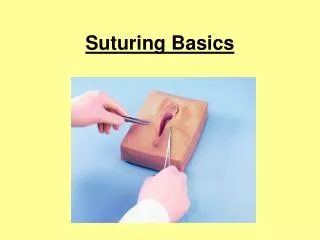
Technique • It is important that the wound is closed in equal ‘bites’ both horizontally and vertically. • The wound edges should be slightly everted.
Technique • Hold the needle (with needle holder/driver) a quarter distance from the blunt end. • The needle should enter the skin with a ¼ inch bite from wound edge at a 90 degree angle.
Technique • Release needle, reach into wound, grasp needle and pull free. (one side of the wound is ‘done’ at a time) • Using forceps, lightly grasp the skin edge and arc needle through opposite edge, inside the wound. • Release the needle, and grasp the protruding needle from the skin and pull through. (leave approx ½ to 1 inch protruding from bite sides)
Technique • Release needle and wrap suture around the needle holder 2 times. • Grasp the ends of the suture material and pull the 2 lines across wound site in opposite directions. (this is called ‘a throw’) • Repeat 3-4 times, but on each throw reverse the order of the wrap. • Cut ends of suture ¼ inch from knot • Do not position knot directly over the wound edge.
Suture Removal • Times will vary according to the location and depth of the wound. However, the average time frame is 7-10 days after application • Face • Body and Scalp • Soles, Palms, Back or Other Joints • Any suture with pus or signs of infections should be removed immediately.
Suture Removal • Using the tweezers, grasp the knot and snip the suture below the knot with the scissors as close as possible to the skin. • Pull the suture line through the tissue • Once all sutures have been removed count the sutures. • The number of sutures needs to match the number indicated in the patient's health record.