Download
1 / 38
620 likes | 1.08k Views
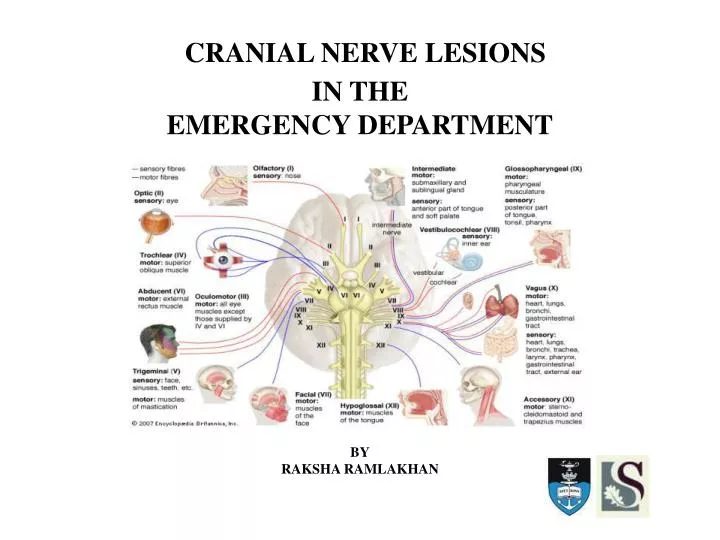
CRANIAL NERVE LESIONS IN THE EMERGENCY DEPARTMENT BY RAKSHA RAMLAKHAN. OVERVIEW. Introduction The cranial nerves 1. Function 2. Anatomy 3. Clinical examination 4. Pathologic features 5. Aetiology Multiple cranial nerve lesions. INTRODUCTION. Challenging for the EP

E N D
CRANIAL NERVE LESIONS IN THE EMERGENCY DEPARTMENT BY RAKSHA RAMLAKHAN
OVERVIEW • Introduction • The cranial nerves 1. Function 2. Anatomy 3. Clinical examination 4. Pathologic features 5. Aetiology • Multiple cranial nerve lesions
INTRODUCTION • Challenging for the EP • Aetiology represent a wide spectrum of pathology • Rapid evaluation for signs and symptoms that are life threatening • Haemorrhage • Infarct • Mass lesion or inflammation with raised intracranial pressure • Consider neoplasms, toxic, metabolic and infectious causes • ABC’S • Frequent re-evaluation if a central cause not excluded • Consultation with neurosurgeon and/or neurologist based on clinical evaluation and imaging
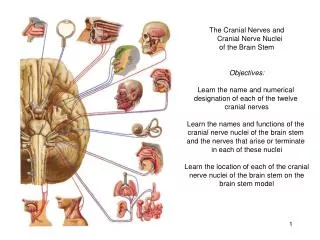
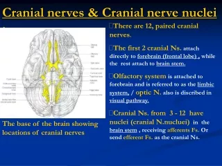
INTRODUCTION - ANATOMY Figure 1:Major nuclei of the cranial nerves Figure 2
THE CRANIAL NERVESCN I – OLFACTORY NERVE FUNCTION: SENSE OF SMELL ANATOMY: - Fibres arise in mucous membrane of nose - Cribriform plate of ethmoid - Synapse in olfactory bulb - Olfactory tract under frontal lobe - Terminates in medial temporal lobe on same side EXAMINATION: - Not routinely tested - Test each nostril separately with bottles containing essences of familiar smells - Examine nasal passages if anosmia present PATHOLOGICAL FEATURES: Unilateral anosmia AETIOLOGY: Trauma – skull fracture or shear injury Tumour – Frontal lobe masses Fig 3
CN II – OPTIC NERVE • FUNCTION : VISION • ANATOMY: - Extends from retina for 5cm - Passes through optic foramen - Joins nerve from other side to form optic chiasm - Fibres from temporal visual fields cross in chiasm whereas those in nasal fields do not - Optic tract to lateral geniculate body - Fibres form optic radiation posterior part of internal capsule - Ends in visual cortex of occipital lobe • CLINICAL EXAMINATION: - Visual acuity - Visual fields - Fundi • PATHOLOGICAL FEATURES: Unilateral/Bilateral visual loss Visual field defects Fig 4 Fig 5
Figure 6: The Visual Fields and Optic Pathways Figure 7: Visual Field Defects
AETIOLOGY: Trauma – traumatic optic neuropathy Tumour – orbital compressive lesion Ischaemic – ischaemic optic neuropathy OPTIC NEURITIS Inflammation of optic nerve complete or partial loss of vision Demyelination Triad of loss of vision, eye pain and impairment of accurate colour vision Causes • Multiple sclerosis • Toxic – ethambutol, chloroquine, nicotine, alcohol • Metabolic – vitamin B12 deficiency • Ischaemia – diabetes, temporal arteritis, atheroma • Familial • Infective – infectious mononucleosis
CN III – OCULOMOTOR NERVE • FUNCTION : MOVES THE EYES - motor fibres to levator palpebrae, superior rectus, medial rectus, inferior rectus, inferior oblique PUPIL CONSTRICTION - parasympathetic fibres to constrictor pupillae and ciliary muscles Fig 8
ANATOMY: Miosis - • Parasympathetic innervation ->Edinger Westphal • Pupil constriction in response to light relayed by optic nerve and tract superior colliculus EW nucleus in midbrain • Efferent motor fibres from oculomotor nucleus • wall of cavernous sinus with CN 4,5a,6 • Iridoconstrictor fibres -ciliary ganglion • Postganglionic fibres innervate iris Figure 9: Pupillary constriction to light
Mydriasis - • Sympathetic innervation to eye • Fibres from hypothalamus to ciliospinal centre and synapse in spinal cord at C8, T1 & T2 • Exit anterior ramus in thoracic trunk & synapse in superior cervical ganglion in neck • Neurones travel with internal carotid artery to eye Figure 10: Oculo Sympathetic Pathway
CLINICAL EXAMINATION: - Pupils – light reflex (direct and consensual) - Accommodation - Eye movements (diplopia and nystagmus) ACCOMMODATION • Neural circuit to visual cortex and back • Involves parasympathetic component of CNIII • Stimulates smooth muscle of ciliary body to contract → lens changes shape • Pupil constricts • Argyll Robertson pupil → absent light reflex with intact accommodation (midbrain lesion)
PATHOLOGICAL FEATURES: Ptosis caused by loss of levator palpebrae function Eye deviated laterally and down Diplopia Dilated non reactive pupil Loss of accommodation Figure 11: Features of CNIII lesion
AETIOLOGY: TRAUMA • Herniation of temporal lobe through tentorial opening ->compression and stretch injury CENTRAL CAUSES • Vascular lesions in brainstem • Tumours • Demyelination PERIPHERAL CAUSES • Compressive lesions -intracranial aneurysms (on posterior comm artery) -tumour -meningitis -nasopharyngeal carcinoma -orbital lesions • Ischaemia/ Infarction -diabetes mellitus -arteritis -migraine -
MEDICAL VS SURGICAL 3rd NERVE PALSY SURGICAL • Post communicating artery aneurysm • Pupillary involvement • Why? Leakage of blood from aneurysm dome -- nerve across outer margin Pupil fibres located very superficially • Cerebral angiography – definitive test • LP – Blood in CSF, inflammation, infection, neoplasm MEDICAL • Pupil sparing • Hallmark of ischaemic lesions - central core of the nerve • Micro vascular disease, insufficiency of vasa nervosa • Frequent in >60yrs and atherosclerotic risk factors • Workup for eg DM,HYPERTENSION (if no evidence of aneurysm) • Spontaneous remission in 6-8 weeks • NSAIDS for pain • Symptomatic Rx
CN - IV TROCHLEAR NERVE • FUNCTION: Motor to superior oblique muscle – intorts ,depresses and abducts globe • ANATOMY: -Nucleus in tegmentum of midbrain -Exits from dorsal aspect -Courses between post cerebral and superior cerebellar arteries before entering cavernous sinus -Enters orbit through superior orbital fissure ,crosses medially over LPS and SRSO • EXAMINATION: Ask patient to turn eye in and then try to look down • PATHOLOGICAL FEATURES: Inability to move eye downward and laterally Diplopia Patients tilt head toward unaffected eye to overcome inward rotation of affected eye • AETIOLOGY: Trauma
A = involved (right) eye is elevated on forward gaze B = extent of elevation is increased with adduction C= extent of elevation is decreased with abduction D = Elevation is increased with head tilting to the affected side E = Elevation is decreased with head tilting in the opposite direction
CN V – TRIGEMINAL NERVE • FUNCTION: Motor to muscles of mastication and tensor tympani Sensory to face, scalp, oral cavity (teeth and tongue) • ANATOMY: • Motor nucleus and sensory nucleus for touch in pons • Proprioceptive nucleus in midbrain • Pain and temperature nucleus descends through medulla to reach upper cervical cord • Cerebellopontine angle temporal lobe in middle cranial fossa • Petrous temporal bonetrigeminal ganglion3 1. Ophthalmic - cavernous sinus with CN3 SOF skin of forehead, cornea and conjunctiva 2. Maxillary -infraorbital foramen skin in middle of face ,mucus membranes of mouth, palate and nasopharynx 3. Mandibular with motor part of nerve foramen ovale skin of lower jaw , mucus membranes of mouth Fig 12
EXAMINATION: -Corneal reflex -Facial sensation -Motor – clench teeth and palpate masseter -Jaw Jerk • PATHOLOGICAL FEATURES: -Partial facial anaesthesia -Episodic facial pain with triggers eg eating and brushing teeth • AETIOLOGY: -Trauma – facial bone fracture Trigeminal neuralgia -Idiopathic, Vascular compression of root -Episodic unilateral facial pain with triggers -Normal findings on head and neck examination and no neurological deficits
-Central causes (pons, medulla and upper cervical cord) vascular tumour syringobulbar -Peripheral (middle fossa) aneurysm tumour chronic meningitis -Trigeminal ganglion (petrous temporal bone) trigeminal neuroma meningioma middle cranial fossa fracture -Cavernous sinus ophthalmic division only with 3,4,6 aneurysm, tumour, thrombosis
CN VI – ABDUCENS NERVE • FUNCTION: Motor supply to lateral rectus muscle (abducts the eye) • ANATOMY: • Leaves brainstem at junction of the pons and medulla, medial to facial nerve. • Runs supant • Subarachnoid spaceemerges from brainstem. • Pons and clivus,dura-between dura and skull. • Tip of petrous temporal bone enter cavernous sinus. • Alongside internal carotid artery. • Enters orbit SOFlateral rectus Fig 13
PATHOLOGICAL FEATURES: Inability to move eye laterally Diplopia on lateral gaze • AETIOLOGY: -Tumour e.g. lesions in cerebellopontine angle -Cavernous sinus lesions e.g. vascular -Elevated intracranial pressure from any cause -Vascular -Metabolic (Wernicke-Korsakoff syndrome) -Subarachnoid space lesions (haemorrhage, infection, inflammation, tumour) -Inflammatory (post viral, demyelinating, giant cell arteritis) Fig 14 note in (A) that the affected (right) eye is adducted at rest - note in (B) that the affected (right) eye cannot abduct
CN VII – FACIAL NERVE • FUNCTION: Motor to muscles of facial expression Parasympathetic stimulation of lacrimal, submandibular and sublingual glands Sensation to anterior two thirds of tongue, ear canal and tympanic membrane Figure 15: Muscles of facial expression
ANATOMY: • Nucleus lies in pons • Leaves pons with CN8 through cerebellopontine angle • Enters facial canal and becomes geniculate ganglion • Gives off nerve to stapedius • Chorda tympani (taste from and two thirds of tongue) joins nerve in facial canal • Leaves skull via stylomastoid foramen • Enters parotid gland and divides to supply muscles of facial expression Fig 16
EXAMINATION: Inspect for facial asymmetry – unilateral drooping of corner of mouth -- smoothing of wrinkled forehead and nasolabial fold Muscle power – look up to wrinkle forehead - shut eyes tightly - smile to compare nasolabial grooves • PATHOLOGICAL FEATURES: • Hemifacial paralysis -LMN (level of nucleus or nerve root) leaves entire side of face paralyzed -UMN (above level of brainstem nucleus) preserves forehead musculature (due to bilateral cortical representation) Figure 17: Left UMN facial weakness
Abnormal taste • Sensory deficit around ear • AETIOLOGY: INFECTION Ramsay Hunt Syndrome • Herepes zoster oticus • unilateral facial paralysis, herpetiformvesiicular eruption, vestibulocochlear dysfunction • Rx similar • Lower incidence of facial recovery and possible sensorineural loss Lyme Disease Bacterial infections
Bells Palsy • (60-70% of acute unilateral facial paralysis) • Idiopathic facial paralysis • ?viral cause – herpes • Abrupt onset LMN paresis progresses over 1-7 days to complete paralysis • Associated s& s – ear pain, decreased tearing, hyperacusis ,impairment of taste • Medical Rx – corticosteriods (1mg/kg per day for 7-10 days) oedema of nerve confined within facial canal causes/contributes to nerve injury • Diabetics have 29% risk of being affected Fig 18 : Bell’s Palsy
UPPER MOTOR NEURON -vascular, tumours TRAUMATIC -Temporal bone fracture with nerve transection -Surgical exploration if evidence -Sudden onset of complete unilateral facial paralysis -Loss of electrical activity -Evidence of displaced fracture involving the facial canal. NEOPLASTIC • -Tumorsof the facial nerve , along course of nerve that invade or compress the nerve. -Progressive -3 weeks -Suspect neoplasticcause if recurrent ipsilateral facial paralysis significant pain, prolonged symptoms, cranial nerve abnormality.
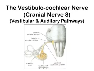
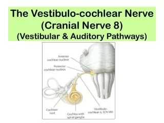
CN VIII – VESTIBULOCOCHLEAR NERVE • FUNCTION: Hearing and balance • ANATOMY: 2 components: -cochlear with afferent fibres for hearing -originate in Organ of Corticochlear nucleus in ponsbilateral transmission to -Medial geniculate bodiessuperior gyrus of temporal lobes -vestibular with afferent fibres for balance -Begin in utricle and semicircular canalsjoin auditory fibres in facial canalenter brainstem at -Cerebellopontine angle.After entering pons-vestibular fibres run widely throughout brainstem and cerebellum Fig 19
EXAMINATION: Hearing test Suspect partial deafness do Rinne’s and Weber’s • PATHOLOGICAL FEATURES: Unilateral hearing loss Tinnitus Vertigo and unsteadiness • AETIOLOGY: -Unilateral nerve deafness tumours eg acoustic neuroma trauma eg petrous temporal bone fracture -Bilateral nerve deafness degeneration toxicity eg aspirin, streptomycin infection eg rubella, syphilis Menieres Disease
CN IX – GLOSSOPHARYNGEAL NERVE & VAGUS NERVES • FUNCTION: General sensation and taste to posterior third of tongue Motor supply to stylopharyngeus • ANATOMY: Nerve fibres from nuclei in medulla form multiple nerve rootlets as they exit the medulla Join to form 9 and 10 CN and also contribute to 11th Emerge from skull through jugular foramen. 9th receives sensory fibres from nasopharynx, pharynx middle and inner ear and post third tongue Also carries secretory fibres to parotid gland Tenth receives sensory fibres from pharynx, larynx & innervates muscles pharynx ,larynx and palate Fig 20
EXAMINATION: Inspect palate and uvula Assess hoarseness and swallowing • PATHOLOGICAL FEATURES: Unilateral 10th nerve palsy –uvula drawn to one side --loss of palatal elevation • AETIOLOGY: -Central causes vascular, tumours, motor neurone disease -Peripheral cause aneurysms at base of skull, tumours, GBS, chronic meningitis
CN XI – ACCESSORY NERVE • FUNCTION: Motor supply to sternocleidomastoid and trapezius muscles • ANATOMY: -Central portion arises in medulla close to nuclei of 9th,10th,12th nerves -Provides motor fibres to vagus -Spinal portion from upper 5 cervical segments I-nnervates trapezuis and SCM • EXAMINATION: Patient shrug shoulders and examiner attempts to push down Turn head to side against resistance of examiners hand • PATHOLOGICAL FEATURES: Downward and lateral rotation of scapula and shoulder drop • AETIOLOGY: Unilateral – trauma, tumours near jugular foramen, polio, syringomyelia Bilateral – motor neurone disease, polio, GBS
CN XII – HYPOGLOSSAL NERVE • FUNCTION: Motor supply to intrinsic and extrinsic muscles of tongue • ANATOMY: Arises from medulla and leaves skull via hypoglossal foramen • EXAMINATION: Inspect tongue – wasting and fasciculations • PATHOLOGICAL FEATURES: Deviation of tongue UMN lesion usually bilateral – tongue deviates towards opposite side NB. Has bilateral UMN innervation LMN lesion – tongue deviates towards side of lesion/weaker affected side - fasciculation wasting and weakness - bilateral causes dysarthria Fig 21
AETIOLOGY: -Bilateral UMN – Vascular, motor neurone disease, tumours -Unilateral LMN – Central – vascular eg thrombosis of vertebral artery, syringobulbia Peripheral – aneurysms, tumours, trauma, meningitis (post fossa) - tumours and lymphadenopathy (upper neck) - Arnold-Chiari malformation -Bilateral LMN GBS, Motor neurone disease polio
MULTIPLE CRANIAL NERVE PALSIES • Anatomical course =>can be affected in groups by single lesions • Syndromes: CAVERNOUS SINUS LESION – unilateral 3,4,5,6 CEREBELLOPONTINE ANGLE LESION –unilateral 5,7,8 JUGULAR FORAMEN LESION – unilateral 9,10,11 PSEUDOBULBAR PALSY • Combined bilateral UMN lesions of 9,10,12 • Long tract signs • Causes – bilateral cerebrovascular disease, MS, Motor neurone disease BULBAR PALSY • Combined bilateral LMN lesions of 9,10,12 • Wasted tongue • Causes – GBS syndrome, brainstem infarction.polio
REFERENCES • Talley NJ, O’Connor S. Clinical Examination. 5th ed.Elsevier;2006 • Rosen P. Emergency Medicine : Concepts and Clinical practice 7th ed. Mosby