Download
1 / 38
380 likes | 639 Views
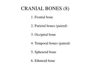
CRANIAL BONES (8). 1. Frontal bone 2. Parietal bones (paired) 3. Occipital bone 4. Temporal bones (paired) 5. Sphenoid bone 6. Ethmoid bone. Frontal. Parietal. Ethmoid. Sphenoid. Temporal. Sphenoid. Sphenoid. Temporal. Temporal. Occipital. P. F. O. P. Frontal. Ethmoid.

E N D
CRANIAL BONES (8) 1. Frontal bone 2. Parietal bones (paired) 3. Occipital bone 4. Temporal bones (paired) 5. Sphenoid bone 6. Ethmoid bone
Frontal Parietal Ethmoid Sphenoid Temporal
Sphenoid Sphenoid Temporal Temporal Occipital
P F O P
Frontal Ethmoid Temporal Sphenoid Parietal (Petrous portion) Temporal (Petrous portion) Occipital
Frontal Sphenoid Sphenoid Perpendicular plate of ethmoid Middle nasal conchae of ethmoid
FACIAL BONES (14) 1. Mandible 2. Maxillae (paired) 3. Palatine bones (paired) 4. Zygomatic bones (paired) 5. Lacrimal bones (paired) 6. Nasal bones (paired) 7. Vomer 8. Inferior Nasal Conchae (paired)
Nasal Lacrimal Zygomatic Maxilla Mandible
Nasal bones Zygomatic Zygomatic Vomer Maxilla Maxilla Inferior nasal conchae Mandible
Maxillae (palatine processes) Vomer Palatine bones (form part of the orbit)
Orbital process of palatine bone
CRANIAL/FACIAL BONES Special Structures
Supraorbital foramen or notch (bilateral) Superior orbital fissure (bilateral) Inferior orbital fissure (bilateral) Infraorbital foramen (bilateral) Mental foramen (bilateral)
Lambdoidal suture Coronal suture Anterior End Posterior End Sagittal suture
Frontal suture Zygomatic process of the temporal bone Squamosal suture Mandibular fossa (Depression where mandible attaches to the temporal bone) Mental foramen
Styloid process Styloid process Mandibularfossa (bilateral) Mastoid process (bilateral)
Squamosal suture Zygomatic bone (facial bone) Zygomatic process of the temporal bone External auditory meatus Mandibular fossa Styloid process Mastoid process
Crista galli Cribiform plate (on both sides, lateral to the Crista galli) Optic foramen (bilateral) Foramen rotundum (bilateral) Sella turcica Foramen ovale (bilateral) Foramen lacerum (bilateral) Foramen spinosum (bilateral)
Lesser wing (sphenoid) - bilateral Greater wing (sphenoid) - bilateral Jugular foramen (bilateral) Internal acoustic meatus (bilateral) Foramen magnum Hypoglossal canal (bilateral)
Foramen lacerum (bilateral) Foramen ovale (bilateral) Carotid canal (bilateral) Stylomastoid foramen (bilateral) Occipital condyle (bilateral) External occipital crest
Carotid canal Jugular foramen (one opening) External occipital protuberance Incisive fossa (may be referred to as a foramen) Stylomastoid foramen Styloid process Mastoid process
Mandibular condyle Coronoid process Mandibular notch Mandibular ramus Alveolus (tooth socket) Mental foramen Mandibular angle Mandibular symphysis
Mandibular condyle Coronoid process Mandibular notch Mandibular foramen (bilateral)
HYOID BONE (Anterior side) Lesser horn Body Superior view (Posterior side) Greater horn
SINUSES Frontal Sphenoid Ethmoid Maxillary From Atlas of the Human Skeleton, Hutchinson, Figure 15, page 16.
VERTEBRAL COLUMN 1. Cervical (7 Vertebrae) Atlas (C1) Axis (C2) Vertebra prominens (C7) 2. Thoracic (12 Vertebrae) 3. Lumbar (5 Vertebrae) 4. Sacrum (1 bone; 5 fused parts) 5. Coccyx (1 bone; 3-5 fused parts)
Atlas (C1) superior view Vertebral foramen Atlas = “yes” bone S Superior articular facets* (Posterior side) Axis = “no” bone Transverse foramen *Attach to occipital condyles Odontoid process or dens of axis (C2) would “fit” here (inferior to this placement) (Anterior side)
Posterior C2 Spinous process Lamina Inferior articular process Pedicle Superior articular facet Transverse process Dens Body (c) Superior view of axis (C2) Figure 7.19c
Typical cervical vertebra superior view Bifid spinous process Inferior articular facet (inferior side of vertebra) Vertebral foramen Transverse foramen Superior articular facet Body
Typical thoracic vertebra lateral view Superior articular process Costal demifacet for head of rib Transverse process (bilateral) Body rib Transverse facet for tubercle of rib Spinous process Costal demifacet for head of rib Inferior notch (becomes intervertebral foramen when vertebrae are “stacked”) Inferior articular process
Typical lumbar vertebra superior view Spinous process Vertebral foramen Lamina Transverse process Vertebral arch Pedicle Body Superior articular process
Sacrum posterior view Superior articular process (facet) Ala Sacral foramen Median sacral crest
Sacrum anterior view Body Ala* Sacral foramen Coccyx *Alae (plural)
Sacrum superior view Superior articular process (facet) Ala Body Coccyx
BONY THORAX • Sternum • Ribs (12 Pairs) • a. Vertebrosternal (true) ribs (1-7) • b. False ribs • 1) Vertebrochondral ribs (8-10) • 2) Vertebral (“Floating”) ribs (11-12)
Sternum Jugular notch Clavicular notch Manubrium Sternal angle Body Xiphisternal joint Xiphoid process
Typical rib superior view Head of rib Neck of rib Costal end Tubercle of rib Shaft of rib (Posterior end - Attaches to vertebra) (Anterior end - Attaches to sternum)
Typical rib inferior view Angle of rib Costal groove Tubercle of rib Neck of rib Head of rib