Download

1 / 66
720 likes | 1.17k Views
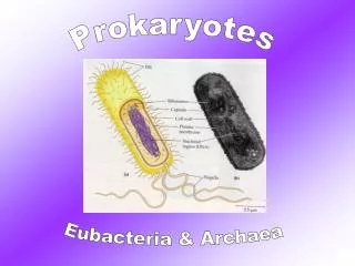
Characterizing and Classifying Prokaryotes. 11. General Characteristics of Prokaryotic Organisms. Prokaryotes Most diverse group of cellular microbes Thrive in various habitats Only a few capable of colonizing humans and causing disease. Figure 11.1 Typical prokaryotic morphologies.

E N D
General Characteristics of Prokaryotic Organisms • Prokaryotes • Most diverse group of cellular microbes • Thrive in various habitats • Only a few capable of colonizing humans and causing disease
General Characteristics of Prokaryotic Organisms • Endospores • Produced by Gram-positive bacteria Bacillus and Clostridium • Each vegetative cell transforms into one endospore • Each endospore germinates to form one vegetative cell • Defensive strategy against unfavorable conditions • Concern to food processors, health care professionals, and governments
Figure 11.2 Locations of endospores. Endospore Endospores
General Characteristics of Prokaryotic Organisms • Reproduction of Prokaryotic Cells • All reproduce asexually • Three main methods • Binary fission (most common) • Snapping division • Budding
Bacterial Growth: Overview PLAY Bacterial Growth: Overview
Figure 11.3 Binary fission. Cell wall Cell replicates its DNA. 1 Cytoplasmic membrane Nucleoid Replicated DNA 2 The cytoplasmic membrane elongates, separating DNA molecules. 3 Cross wall forms; membrane invaginates. 4 Cross wall forms completely. 5 Daughter cells may separate.
Figure 11.4 Snapping division, a variation of binary fission. Older, outer portion of cell wall Newer, inner portion of cell wall Rupture of older, outer wall Hinge
Figure 11.5 Spores of actinomycetes. Filamentous vegetative cells Spores
General Characteristics of Prokaryotic Organisms • Reproduction of Prokaryotic Cells • Epulopiscium and its relatives have unique method of reproduction • Live offspring emerge from the body of the dead mother cell (viviparity) • First noted case of viviparous behavior in prokaryotic world
General Characteristics of Prokaryotic Organisms • Arrangements of Prokaryotic Cells • Result from two aspects of division during binary fission • Planes in which cells divide • Separation of daughter cells
Figure 11.7 Arrangements of cocci. Plane of division Diplococci Streptococci Tetrads Sarcinae Staphylococci
Figure 11.8 Arrangements of bacilli. Single bacillus Diplobacilli Streptobacilli Palisade V-shape
Modern Prokaryotic Classification • Currently based on genetic relatedness of rRNA sequences • Three domains • Archaea • Bacteria • Eukarya
Figure 11.9 Prokaryotic taxonomy. PHYLUM CHLOROFLEXI (green nonsulfur) BACTERIA Thermophilic bacteria PHYLUM DEINOCOCCUS-THERMUS Deeply branching bacteria GRAM-NEGATIVE BACTERIA PHYLUM PROTEOBACTERIA PHYLUM AQUIFICAE Purple nonsulfur () Nitrifying () Rickettsias () Nitrogen fixing () Myxobacteria () PHYLUM CHLOROBI (green sulfur) Campylobacteria () Gammaproteobacteria () PHYLUM CYANOBACTERIA Neisserias () PHYLUM FIBROBACTERES PHYLUM BACTEROIDETES PHYLUM CHLAMYDIAE ARCHAEA PHYLUM SPIROCHAETES PHYLUM PLANCTOMYCETES PHYLUM EURYARCHAEOTA Methanogens Low G+C Gram-positive PHYLUM CRENARCHAEOTA PHYLUM FIRMICUTES PHYLUM FUSOBACTERIA Halophiles Clostridia Mycoplasmas Selenomonas Bacilli, lactobacili, cocci Streptomyces Atopobium Corynebacterium Mycobacterium Nocardia Thermophilic archaea Arthrobacter PHYLUM ACTINOBACTERIA GRAM-POSITIVE BACTERIAHigh G+C Gram-positive
Survey of Archaea • Common features • Lack true peptidoglycan • Cell membrane lipids have branched hydrocarbon chains • AUG codon codes for methionine • Two phyla: Crenarchaeota, Euryarchaeota • Reproduce by binary fission, budding, or fragmentation • Are cocci, bacilli, spirals, or pleomorphic • Not known to cause disease
Survey of Archaea • Extremophiles • Require extreme conditions to survive • Temperature, pH, and/or salinity • Prominent members are thermophiles and halophiles
Survey of Archaea • Extremophiles • Thermophiles • DNA, RNA, cytoplasmic membranes, and proteins do not function properly below 45ºC • Hyperthermophiles – require temperatures over 80ºC • Two representative genera • Geogemma • Pyrodictium
Figure 11.11 Some hyperthermophilic archaea live in hot springs.
Survey of Archaea • Extremophiles • Halophiles • Inhabit extremely saline habitats • Depend on greater than 9% NaCl to maintain integrity of cell walls • Many contain red or orange pigments • May protect from sunlight • Most studied – Halobacterium salinarium
Figure 11.12 The habitat of halophiles: highly saline water.
Survey of Archaea • Methanogens • Largest group of archaea • Convert carbon dioxide, hydrogen gas, and organic acids to methane gas • Convert organic wastes in pond, lake, and ocean sediments to methane • Some live in colons of animals • One of primary sources of environmental methane • Have produced ~10 trillion tons of methane that is buried in mud on ocean floor
Survey of Bacteria • Deeply Branching and Phototrophic Bacteria • Deeply branching bacteria • Scientists believe these organisms are similar to earliest bacteria • Autotrophic • Live in habitats similar to those thought to exist on early Earth • Aquifex • Considered to represent earliest branch of bacteria • Deinococcus • Has outer membrane similar to Gram-negatives, but stains Gram-positive
Survey of Bacteria • Deeply Branching and Phototrophic Bacteria • Phototrophic bacteria • Phototrophs that contain photosynthetic lamellae • Autotrophic • Divided into five groups based on pigments and source of electrons for photosynthesis • Blue-green bacteria (cyanobacteria) • Green sulfur bacteria • Green nonsulfur bacteria • Purple sulfur bacteria • Purple nonsulfur bacteria
Figure 11.13 Examples of cyanobacteria with different growth habits. Vegetative cell Heterocyst Akinete Sheath (glycocalyx)
Figure 11.14 Deposits of sulfur within purple sulfur bacteria.
Photosynthesis: Comparing Prokaryotes and Eukaryotes Photosynthesis: Comparing Prokaryotesand Eukaryotes PLAY
Survey of Bacteria • Low G + C Gram-Positive Bacteria • Clostridia • Rod-shaped, obligate anaerobes • Important in medicine and industry • Microbes related to Clostridium include Epulopiscium, sulfate-reducing microbes, and Selenomonas
Survey of Bacteria • Low G + C Gram-Positive Bacteria • Mycoplasmas • Facultative or obligate anaerobes • Lack cell walls • Smallest free-living cells • Colonize mucous membranes of the respiratory and urinary tracts
Figure 11.15 The distinctive "fried egg" appearance of Mycoplasma colonies.
Survey of Bacteria • Low G + C Gram-Positive Bacteria • Other low G + C bacilli and cocci • Bacillus • Many common in soil • Bacillus thuringiensis toxin used by farmers and gardeners as an insecticide • Bacillus anthracis causes anthrax
Figure 11.16 Crystals of Bt toxin, produced by the endospore-forming Bacillus thuringiensis. Bacillus thuringiensis Bt toxin
Survey of Bacteria • Low G + C Gram-Positive Bacteria • Other low G + C bacilli and cocci • Listeria • Contaminates milk and meat products • Capable of reproducing under refrigeration • Can cross the placenta in pregnant women • Lactobacillus • Grows in the body but rarely causes disease • Used in the production of various foods
Survey of Bacteria • Low G + C Gram-Positive Bacteria • Other low G + C bacilli and cocci • Streptococcus and Enterococcus • Cause numerous diseases • Various strains of multi-drug-resistant streptococci • Staphylococcus • One of the most common inhabitants of humans • Produce toxins and enzymes that contribute to disease
Survey of Bacteria • High G + C Gram-Positive Bacteria • Corynebacterium • Pleomorphic aerobes and facultative anaerobes • Produces metachromatic granules • Mycobacterium • Aerobic rods that sometimes form filaments • Slow growth partly due to mycolic acid in its cell walls • Actinomycetes • Form branching filaments resembling fungi • Important genera include Actinomyces, Nocardia, Streptomyces
Figure 11.17 The branching filaments of actinomycetes. Spores
Survey of Bacteria • Gram-Negative Proteobacteria • Largest and most diverse group of bacteria • Five classes of proteobacteria • Alphaproteobacteria • Betaproteobacteria • Gammaproteobacteria • Deltaproteobacteria • Epsilonproteobacteria
Survey of Bacteria • Gram-Negative Proteobacteria • Alphaproteobacteria • Nitrogen fixers • Two genera important to agriculture • Grow in association with the roots of plants • Azospirillum • Rhizobium
Figure 11.18 A prostheca. Flagellum Prostheca
Survey of Bacteria • Gram-Negative Proteobacteria • Alphaproteobacteria • Nitrifying bacteria • Oxidation of nitrogenous compounds provides electrons • Important in the environment and agriculture • Nitrobacter • Purple nonsulfur phototrophs • Grow at the bottom of lakes and ponds
Survey of Bacteria • Gram-Negative Proteobacteria • Alphaproteobacteria • Pathogenic alphaproteobacteria • Rickettsia • Transmitted through bite of an arthropod • Cause several human diseases • Brucella • Causes brucellosis
Survey of Bacteria • Gram-Negative Proteobacteria • Alphaproteobacteria • Other alphaproteobacteria • Acetobacter • Gluconobacter • Caulobacter
Figure 11.20 Growth and reproduction of Caulobacter. Rock or other substrate 1 Prostheca Flagellum 2 Swarmer cell Cell doubles in size 3 4a or 4b Rosette