Download

1 / 22
220 likes | 418 Views
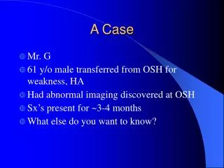
FBC Case A. Kelly Jen MyLinh. The case…. A 61 year old man presented with a 3 week history of generalised weakness & increasing dyspnoea on exertion. He has a history of alcoholism, has been divorced for 2 years and lives alone. He has not worked since being retrenched at the age of 55.

E N D
FBC Case A Kelly Jen MyLinh
The case… A 61 year old man presented with a 3 week history of generalised weakness & increasing dyspnoea on exertion. He has a history of alcoholism, has been divorced for 2 years and lives alone. He has not worked since being retrenched at the age of 55. On examination pallor and scleral icterus were noted. There was clinical evidence of chronic alcoholic liver disease with portal hypertension. The spleen was palpable (2 cm)
Blood film – marked anisocytosis (oval macrocytes +++) and poikilocytes (tear drop & fragmented cells ++). Red cells normochromic. Neutropenia with marked neutrophil hypersegmentation. Thrombocytopenia.
Chronic alcoholic abuse/alcoholic liver disease • Almost all alcohol metabolised by the liver • Directly hepatotoxic • 3 stages of alcoholic liver disease • Hepatic steatosis • Alcoholic hepatitis • Alcoholic cirrhosis
Hematologic changes in liver disease • Anaemia • Leukopenia • Thrombocytopenia • Often with splenomegaly in portal hypertension
Other changes in liver disease • Late stage liver disease… • Liver starts out yellow, fatty and enlarged • As disease progresses, atrophies • Brown • Shrunken • No longer fatty • Biochem… • Elevated serum transaminase • Hyperbilirubinaemia • Hypoproteinaemia • anaemia
What is LD? • Intracellular enzyme widely distributed in all tissues of the body. • Catalyzes the conversion of lactate pyruvate • LD released from cells when damaged
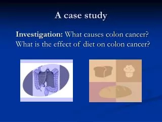
What does raised LD mean? Associated with tissue damage diseases: • AMI ( 36-55hrs after) • CHF • Liver disease (e.g. cirrhosis, alcoholism) • Megaloblastic & pernicious anemia's • RBC destruction ( Hb)
What are Haptoglobins (Hp)? • 2 - globulin that binds free Hb in bloodstream. Results in removal of the complex from circulation • Primary physiologic fn is the preservation of Fe USE: Indicator Chronic Hemolysis Reflects: Hb
What happens when Haptoglobins are reduced? • Hp Decreased or absent in: • Hemogloinemia • Intramedullary Hemolysis (e.g. Megaloblastic anemia) • Acute or Chronic liver disease • [ ] Hp is inversely related to degree of Hemolysis and duration of hemolytic episode
Ferritin • Complex of ferric hydroxide and protein • Reflection of body iron stores • Reliable indicator of Total body iron status USE: Diagnosis of iron deficiency or Iron Excess
What does raised Ferritin reflect? • Acute & chronic liver disease • Alcoholism ( during abstinence) • Malignancies • Infection • Inflammation • AMI • Anemia's other than Iron deficiency • E.g.. Megaloblastic anemia
Serum Folate 0.7 (7-45) nmol/L Explain why folate deficiency is likely in this case?
FOLATE • Absorption of folate occurs in the jejunum • The principal storage site is in the liver • distribution depends mostly on an enterohepatic recirculation, in which folate in a methylated form is reabsorbed from bile into the serum. • After folate enters most tissues, including erythrocytes, it remains throughout the life span of the cell.
CAUSES: inadequate dietary intake malabsorption (e.g. jejunal disease) increased demands (e.g. pregnancy, infancy, leukemia) Drug induced (e.g. anticonvulsants, oral contraceptives, MTX and alcohol). FOLATEDEFICIENCY
Folate Deficiency and Alcohol • Alcohol ingestion interferes with intermediate metabolism of folate, its intestinal absorption and enterohepatic salvage • interfering with the release of folate from the liver into the bile -> a rapid DECREASE in the SERUM FOLATE LEVEL).
Do the results support the diagnosis of megaloblastic anaemia?
Megaloblastic Anaemia • Folic acid deficiency and Vitamin B12 deficiency anaemia are the two most common examples. • These deficiencies cause defective DNA synthesis, whereas RNA synthesis and synthesis of cytoplasmic components unaffected -> (cytoplasmic maturity>nuclear maturity) -> megaloblast in marrow.
…continued • Blood films show oval macrocytes and hypersegmented neutrophil nuclei (with 6 lobes) • In severe cases, WCC and platelet count also fall (pancytopenia). The bone marrow show characteristic megaloblastic erythroblasts. • Dyspoiesis increases intermedullary cell death (ineffective erythropoeisis) with resultant indirect hyperbilirubinemia and hyperuricaemia
Glossitis (sore pale smooth tongue) Altered bowel habit Mild jaundice Insidious onset Pallor Loss of appetite Bilateral peripheral neuropathy (vit B12 deficient only) Tiredness Headaches In severe anaemic pxs may present with CHF Clinical features
Yes, the results do support the diagnosis!! • Blood film: marked anisocytosis (oval macrocytes+++), poikilocytes (tear drop and fragmented cells++), neutropenia with marked neutrophil hypersegmentation and thrombocytopenia • Clinical symptoms: scleral icterus, pallor, generalised weakness and increasing dyspnoea on exertion • Biochemistry: low serum folate and red cell folate